Tissue Sampling • Gomti Nagar
Image-Guided Biopsy in Lucknow
A biopsy takes a small piece of tissue from a lump or organ so a pathologist can examine it. Imaging guides the needle to the right place, which makes the sample safer and more useful.
- Vijayant Khand, Gomti Nagar, Lucknow
- Mon to Sat 9 AM to 8 PM, Sun 10 AM to 2 PM
- Sample
- Thin cores of tissue
- Numbing
- Local anaesthetic is usual
- Before
- Tell us about blood thinners
- Report
- Goes to your referring doctor
What a biopsy is
A biopsy is the removal of a small sample of tissue so that it can be examined under a microscope. It is the most direct way to find out what an abnormal area really is, because a scan can show a lump but cannot always name it.
This page is about needle biopsy. In a core biopsy, a hollow needle takes a thin thread of tissue, no thicker than a pencil lead. Surgery removes larger samples or the whole lump, and that is a separate decision. Loose cells alone are collected by FNAC, which uses a finer needle.
Why a doctor advises a biopsy
A doctor advises a biopsy when a scan or an examination finds something that cannot be named without looking at the tissue. Being asked to have one does not mean that cancer has been found. The usual reasons are:
- A lump or shadow of unknown cause on a scan, such as a mass in the liver or an enlarged lymph node.
- To tell cancer from infection or inflammation, which can look alike on images.
- To name the exact type of a tumour, which decides the choice of treatment.
- To look at a whole organ, for example when liver or kidney tests stay abnormal without a clear cause.
A biopsy can also show that an abnormal area is harmless. That can end the worry and spare you treatment you do not need.
How imaging guides the needle
Ultrasound is the usual guide for the liver, thyroid, lymph nodes, kidneys and lumps in soft tissue. It shows the needle as a bright line in real time, so the doctor can place the tip inside the target and keep clear of nearby vessels. The page on ultrasound-guided biopsy explains how that works.
Some targets cannot be seen well on ultrasound, for example those behind gas-filled bowel or deep in the lung. The doctor then advises another route or another method, and the team can look at your images.
Whether a biopsy is done here, and who does it, is confirmed only after the team has read your referral, scans and blood reports. The team will tell you when you book.
Risks and limits of a needle biopsy
Bleeding is the main risk. Most people have only a small bruise, but bleeding can also happen inside an organ, where it cannot be seen. This is why the team checks your blood counts and clotting tests and asks about every blood thinner you take.
Other risks are infection, soreness at the site and injury to a structure beside the target. The doctor chooses a path that avoids vessels and organs on the images, and tells you the risks for your site before you consent.
A biopsy also has a limit: the report may not settle the question. If so, your doctor discusses a repeat sample or a different test.
What happens to the sample
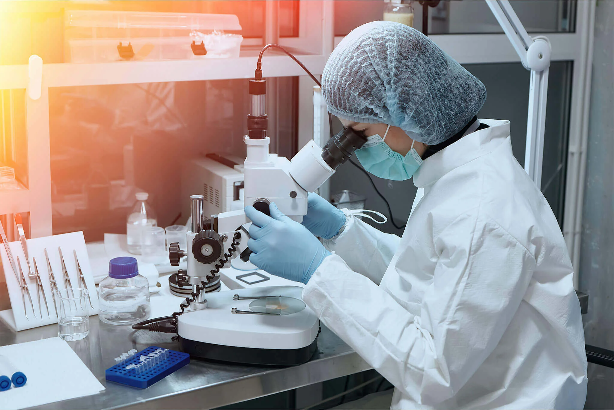
The tissue is placed in a preservative, labelled and sent to a pathology laboratory. A pathologist, a doctor who studies tissue, cuts it into thin slices, stains them and examines them under a microscope. Some samples need extra stains, which takes longer.
The report goes to the doctor who referred you. Please discuss it with them, because they know your scans, symptoms and history, and the same words can mean different things in different people. The team will tell you when to expect it. Blood tests before the procedure can be done at our pathology lab.
Where a needle biopsy is commonly taken
These are examples, not a list of what is done here. Your referral decides the target.
-
Liver
A mass, or the whole liver when tests stay abnormal.
-
Lymph node
Glands in the neck, armpit, groin or abdomen.
-
Thyroid
Nodules that FNAC could not settle.
-
Soft tissue
Lumps in muscle or under the skin.
-
Kidney
A mass, or the kidney itself when kidney disease is unexplained.
-
Handled within our breast care service.
What happens on the day
-
1
Review
The team reads your referral, scans and blood tests, and confirms the target, the method and where the biopsy will be done. You hear the benefits, risks and alternatives.
-
2
Consent and positioning
You sign consent. You then lie in the position that gives the safest path to the target, for example on your back or your side.
-
3
Numbing
The skin is cleaned, and the tissue along the needle path is numbed with local anaesthetic. This stings briefly.
-
4
Sampling
Under imaging, a hollow needle is passed to the target. A spring-loaded device gives a loud click as it takes each core. Several cores are usually taken.
-
5
Observation
Pressure is held on the site and a dressing goes on. You rest for the time the team advises, which depends on the site. Liver biopsies need longer.
-
6
Sample to the lab
The cores are labelled and sent to the laboratory. The report goes to your referring doctor.
Preparation and aftercare
Before your visit
- Have the blood tests the team asks for, usually a blood count and a clotting test.
- Tell the team and your doctor about every blood thinner and painkiller you take, including aspirin, clopidogrel, warfarin and ibuprofen. Do not stop any of them yourself.
- Fasting is needed for some sites, such as the liver or abdomen, and not for others. The team confirms this when you book.
- If you have diabetes and are asked to fast, ask your doctor how to take your insulin or tablets that day.
- Arrange for a relative or friend to come with you and travel home with you.
Please bring
- Your doctor's prescription or referral
- Earlier scans, with reports and images
- Recent blood test reports
- Earlier biopsy or pathology reports
- A list of your medicines
After your visit
- Take the rest of the day easy, and avoid heavy lifting and hard exercise for as long as the team advises.
- Keep the dressing clean and dry. A small bruise and a sore spot are common.
- If your doctor paused a medicine, ask when to restart it. Do not restart it on your own.
- Book a visit with the referring doctor to discuss the report.
When to get urgent help after a biopsy
Most people have no problems, but bleeding and infection can happen. Go to the nearest emergency department at once if you faint, have severe pain or cannot stop bleeding. Contact your doctor or the centre the same day if you have any of these:
- Bleeding that soaks the dressing or does not stop after ten minutes of firm pressure.
- A swelling at the site that grows or feels tense and hard.
- Pain that becomes severe or keeps getting worse, including pain in the abdomen or shoulder after a liver biopsy.
- Dizziness, fainting, a racing heartbeat or cold, clammy skin, which can mean bleeding inside.
- Fever or chills, or redness and pus at the site.
- Heavy blood in the urine after a kidney biopsy, or blood that does not clear.
Image-Guided Biopsy: common questions
How is a biopsy different from FNAC?
FNAC draws loose cells through a very thin needle. A biopsy takes a thread of tissue with a wider needle, so the pathologist can see how the cells are arranged. That gives more detail and is often needed to name a tumour. Your doctor chooses between them, and sometimes FNAC comes first.
Does a biopsy mean I have cancer?
No. A biopsy is advised when the cause of an abnormal area is not clear. Many biopsies show infection, inflammation or a harmless growth. The test exists to find out, so that cancer is not missed and harmless findings are not over-treated.
Does a biopsy hurt?
The numbing injection stings for a few seconds. After that you feel pressure and hear a click from the device, but sharp pain is not expected. Tell the doctor if you feel pain, and more anaesthetic can be given. The site may be sore for a day or two.
Should I stop my blood thinner before a biopsy?
Not on your own. Stopping can be dangerous for people with heart stents or a past stroke, for example. Tell the team what you take and why, and ask the doctor who prescribed it. Together they judge whether a pause is safe, for how long, and when to restart.
When will I get the biopsy result?
The time depends on the tissue and on the tests the pathologist needs, so the team will tell you when you book. The report goes to your referring doctor. Ask them to go through it with you.
Can a biopsy result be unclear?
Yes. The needle may miss the abnormal part, or the cores may be too small to decide. Sometimes the pathologist sees something that needs extra stains or tests first. Your doctor may then advise a repeat biopsy or a different test.
Do I need someone with me on the day?
It is wise to bring a relative or friend. They can listen to the instructions with you and see you home. If you feel faint or sore afterwards, do not travel alone or drive yourself.
Where can I get an image-guided biopsy near Gomti Nagar?
The centre is at 4/12, Vijayant Khand, near Kathauta Chauraha, Gomti Nagar. Send your prescription on WhatsApp at +91 70809 82920 or call 0522-3180995. The team confirms whether the biopsy can be done at the centre or elsewhere, and how to prepare.
Related services
Tests and care that often go together
Appointments
Ask about Image-Guided Biopsy in Gomti Nagar, Lucknow
Send your prescription and any earlier reports on WhatsApp or call the centre. The team reviews them and tells you the next step.
4/12, Vijayant Khand, Gomti Nagar · 0522-3180995