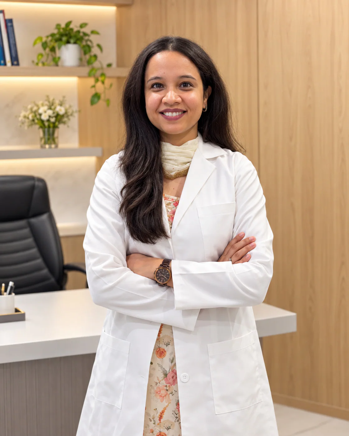
Dr. Pranjali Joshi
Breast Radiologist
- MBBS (KGMU)
- MD Radiodiagnosis (Dr. RMLIMS)
- PDCC Breast Imaging (AIIMS Rishikesh)
Dr. Pranjali Joshi is the breast radiologist at the centre, with an MD in Radiology and a PDCC in Breast Imaging.
Read her profile4/12, Vijayant Khand, Gomti Nagar, Lucknow Mon–Sat 9 AM–8 PM · Sun 10 AM–2 PM
Advance Breast Care • Dense Breasts
Dense breast imaging starts with a mammogram and may add 3D mammography or a breast ultrasound. Dense breasts are common, cannot be felt, and can hide small changes.
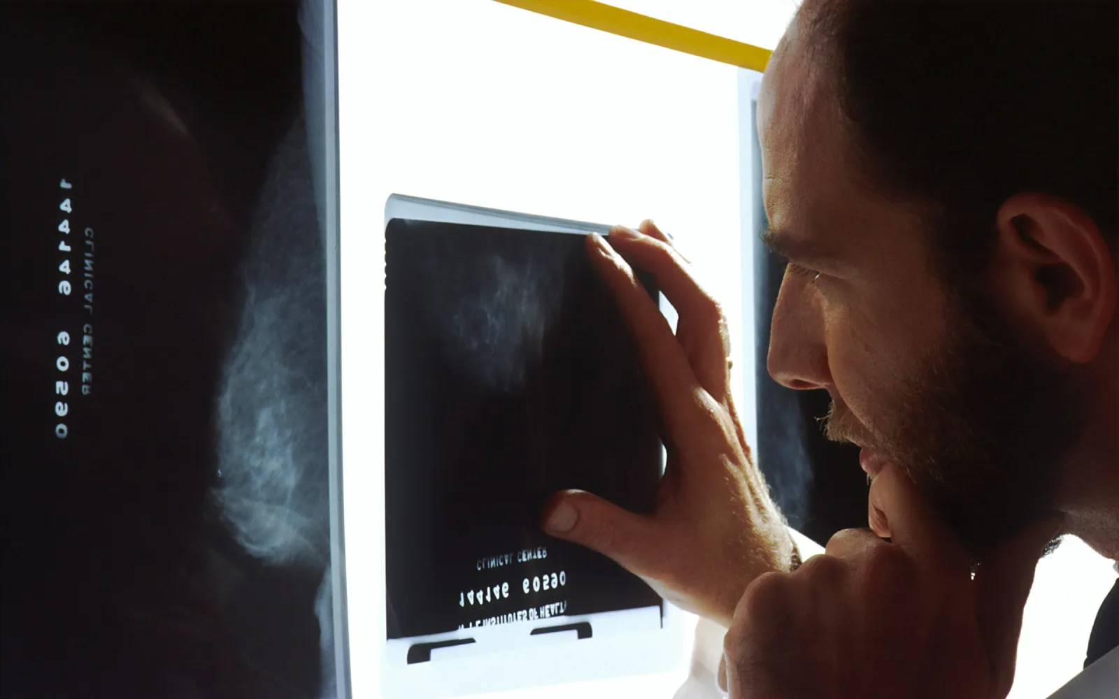
Breast density describes how much of the breast is gland and fibrous tissue compared with fat, as seen on a mammogram. Dense breasts have more of the first and less of the second. It has nothing to do with the size of the breasts or how firm they feel.
Your mammogram report may give a density category. Radiologists use four: almost entirely fatty, scattered areas of dense tissue, heterogeneously dense and extremely dense. The last two are what people usually call dense breasts.
Density is normal. It is more common in younger women and often lessens with age and once periods have stopped, though not in every woman. Dense breasts are also linked with a somewhat higher chance of breast cancer over a lifetime. Density alone is not a diagnosis, and it is not a cause for alarm.
Dense tissue appears white on a mammogram, and so do many small changes, so a small change can be lost against white tissue. Fatty tissue appears dark, which is why a change stands out clearly in a fatty breast.
This does not make the mammogram useless. It still shows many changes, including the tiny calcium specks that a breast ultrasound shows poorly. It means the picture is harder to read and a small change can be missed. That is the reason extra imaging is sometimes added.
Whether you need anything extra depends on your age, your family history, any symptoms and what the mammogram shows. Many women with dense breasts need only the mammogram. When more is advised, it is usually one of these:
These extras are a doctor's decision. Having dense breasts does not mean you will be sent for all of them.
The word itself is no reason to hurry. Ask your doctor whether extra imaging makes sense for you, and keep to the screening schedule your doctor sets.
Two points matter. In dense breasts a normal mammogram is a little less reassuring than in fatty ones, so notice how your breasts look and feel. And if you can feel a lump, a normal mammogram does not settle it: you still need an examination and usually an ultrasound. See breast lump evaluation.
Message or call the centre. Mention that an earlier report said your breasts were dense, and send the prescription and the report.
You have a 2D or 3D mammogram, depending on what your doctor asked for. Each breast is pressed briefly between two plates.
The radiologist reads the pictures, notes the density and compares them with earlier mammograms.
A breast ultrasound may be advised at the same visit or a later one. The team will tell you which, when you book or on the day.
The report usually states the density category and any advice. Take it to your doctor to decide what, if anything, to add.
Before your visit
Please bring
After your visit
A normal mammogram does not rule out a change that you notice yourself. See a doctor soon if you find:
Dr. Pranjali Joshi
Breast Radiologist
Dr. Pranjali Joshi is the breast radiologist at the centre, with an MD in Radiology and a PDCC in Breast Imaging.
Read her profileNo. Dense breasts are a common, normal variation. Density describes how the breast looks on a mammogram and is not a disease. It can make small changes harder to see, which is why doctors pay attention to it.
Only imaging can tell you, usually a mammogram report. You cannot tell by touch, by breast size or by how firm your breasts feel. If your report does not mention density, ask the radiologist or your doctor.
No. Whether to add one depends on your age, family history, symptoms and the mammogram findings. Many women with dense breasts have only a mammogram. Your doctor may suggest an ultrasound if an area needs a closer look.
It can help, because separating the layers lessens the effect of overlapping tissue. It does not turn dense breasts into fatty ones, and for some women a breast ultrasound is still added. Your doctor will advise.
Yes. A mammogram still shows many changes, including tiny calcium specks that other tests find hard to see. It is read with the knowledge that dense tissue can hide small changes, which is why extra imaging is sometimes added.
Not always. Density often lessens with age and once periods have stopped, although not in every woman, and hormone treatment can affect it. Your next mammogram report will show the current category.
We are at 4/12, Vijayant Khand, near Kathauta Chauraha, and we do mammography, 3D mammography and breast ultrasound there. Message the team on WhatsApp at +91 70809 82920 or call 0522-3180995, and send your earlier report so the right combination can be booked.
Related services
The mammogram taken in thin layers.
Learn more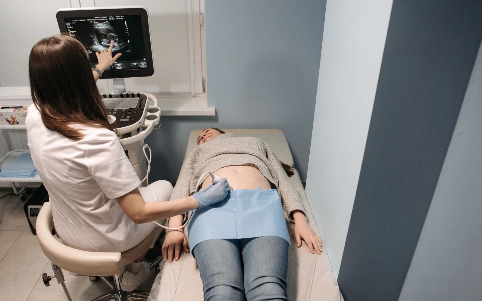
The radiation-free scan often added for dense tissue.
Learn more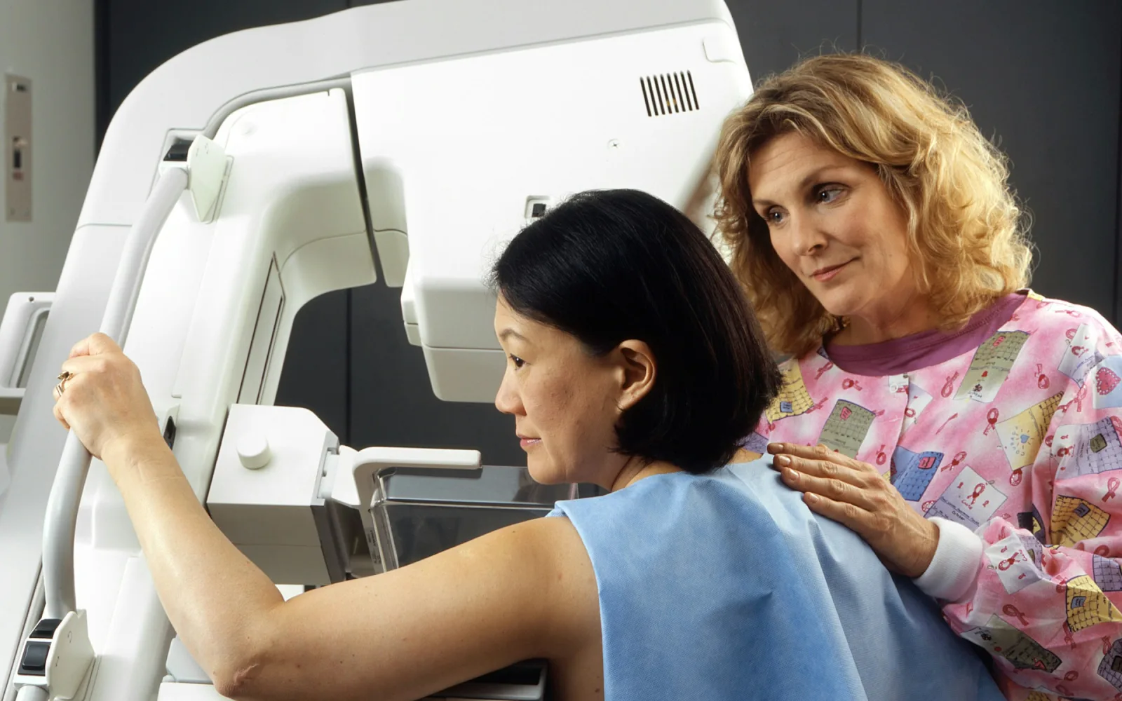
Screening and diagnostic mammograms.
Learn more
Checking for breast cancer before symptoms, usually with a mammogram.
Learn moreAppointments
Message us on WhatsApp or call the centre. Tell us the test you need and your preferred day, and the team will confirm the details.
4/12, Vijayant Khand, Gomti Nagar · 0522-3180995