Dr. Akanksha Bajpai
Radiologist
- MBBS, MD
- Ex. PGI Chandigarh
- Ex Consultant, Fortis Hospital, Mohali
Her profile supports diagnostic imaging, ultrasound, radiology and patient-focused interpretation.
Read her profile4/12, Vijayant Khand, Gomti Nagar, Lucknow Mon–Sat 9 AM–8 PM · Sun 10 AM–2 PM
Joint and Soft Tissue Scans • Gomti Nagar
A joint ultrasound shows tendons, ligaments, muscle, fluid and lumps live on screen, and can be done while you move the joint. No radiation and no fasting, at our centre in Vijayant Khand.
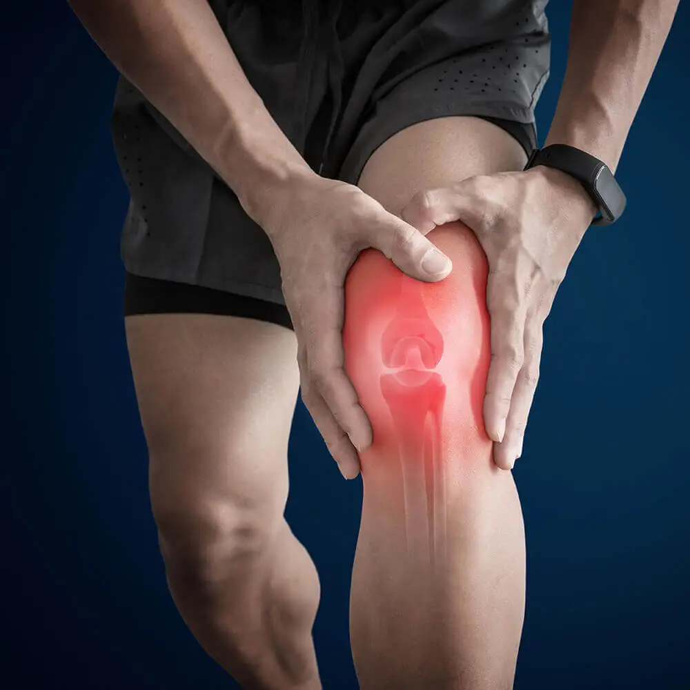
It shows the soft parts around a joint: tendons, which attach muscle to bone, and ligaments, which hold bones together. It also shows muscles, the fluid-filled cushions called bursae, fluid inside the joint, and lumps under the skin. It is clearest for structures close to the surface.
The scan can show a tendon that is swollen, thickened, partly torn or fully torn. It can show fluid in the knee or hip, a swollen bursa, a Baker's cyst behind the knee, a ganglion on the wrist, or a lipoma. It can also pick up a wooden splinter or thorn that an X-ray can miss.
The radiologist can compare with your other side. Colour Doppler can show the extra blood flow in inflamed tissue when that helps.
Doctors ask for it when the question is about soft tissue. Here are the usual problems by area.
A scan is for looking. An injection into a joint or tendon under ultrasound guidance is a separate procedure, covered on our pain injection page, and needs its own medical advice and booking.
Ultrasound can watch a joint work. The radiologist may ask you to lift your arm, bend your knee or straighten your fingers while the probe stays on the area. A shoulder tendon that catches against bone as you raise the arm, a tendon that snaps over a bony edge, or a lump that moves with a muscle may only show this way.
The movements are gentle and kept within what you can manage. Tell the radiologist when something hurts. The sore spot, and the position that brings it on, are both useful. Moving the joint is used where it helps. It is not part of every scan.
The three tests see different things. An X-ray shows bone: fractures, wear in a joint, alignment and calcium deposits. Ultrasound shows soft tissue near the surface: tendons, fluid, lumps and bursae. MRI shows deeper structures, such as menisci, cruciate ligaments and cartilage, in more detail than ultrasound can.
Doctors often start with an X-ray after an injury or for pain in an older joint. They add ultrasound when the question is about a tendon, fluid or a lump. Our centre has no MRI scanner. If your doctor advises one, ask where to have it done. Our team can review the images later, alongside an ultrasound if you need one.
Send the prescription and say which joint and which side hurts. The team confirms the scan and tells you what to wear.
You roll up a sleeve or trouser leg, or change into a gown for the shoulder or hip. You sit or lie so the joint can be reached and moved.
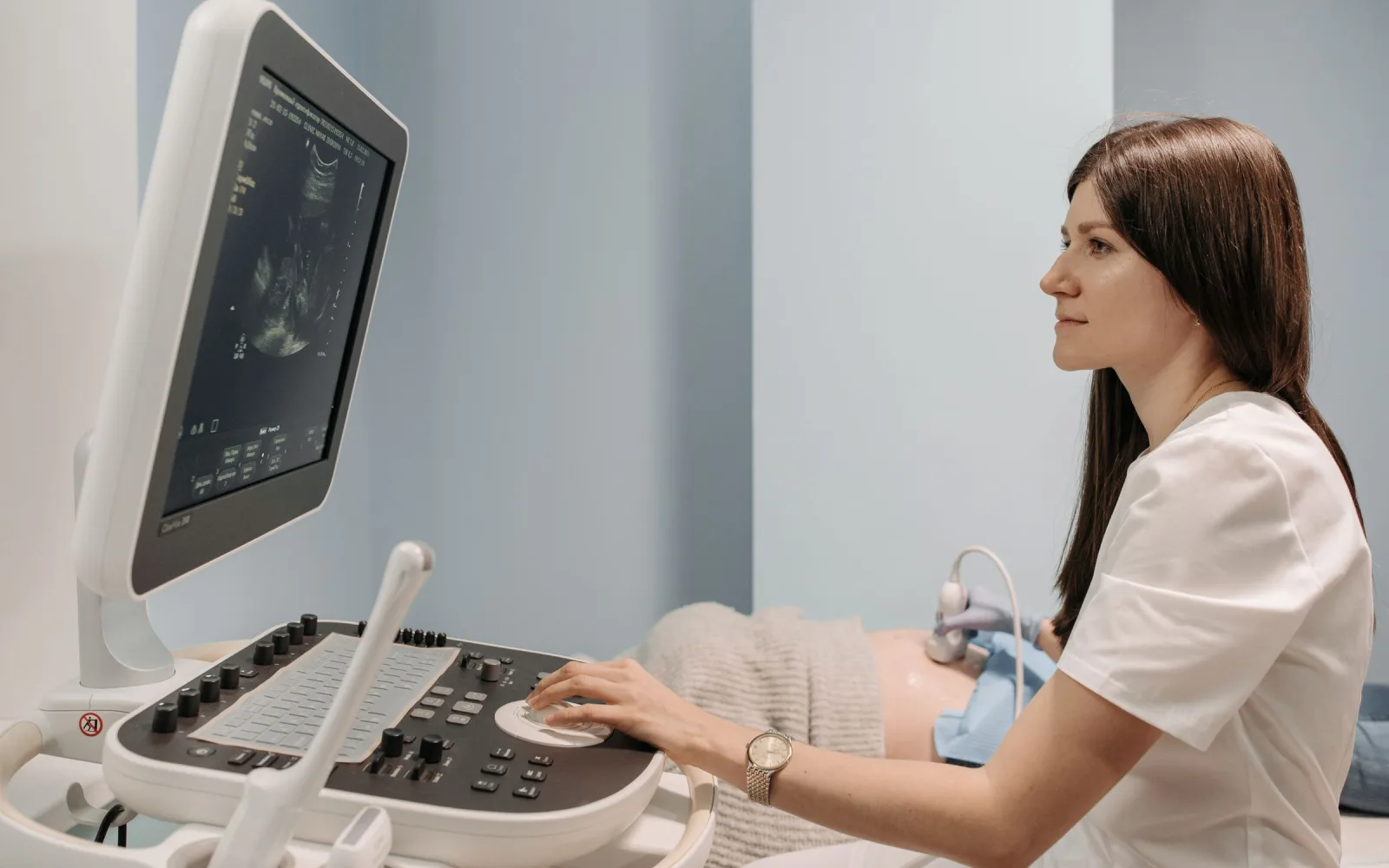
The radiologist spreads warm gel over the joint and glides the probe along the tendons. Pressure is light, though pressing on a sore spot can hurt for a moment. Say when it does.
The radiologist may ask you to move the joint in a set way while watching the screen. They may also scan the other side for comparison.
A written report follows. The team will tell you when and how it reaches you.
Take the report, with any X-rays, to the orthopaedic doctor, physician or physiotherapist who sent you. They decide on rest, exercises, medicines, an injection or further tests.
Before your visit
Please bring
After your visit
Go to a hospital emergency department if you have:
Dr. Akanksha Bajpai
Radiologist
Her profile supports diagnostic imaging, ultrasound, radiology and patient-focused interpretation.
Read her profileNot reliably. These structures lie deep inside the knee, where sound waves do not reach well. Ultrasound is better for tendons, fluid and the structures near the surface. For a suspected meniscus or ligament tear, your doctor will usually advise an MRI, which we do not offer.
It is useful for shoulder pain. It can show a rotator cuff tear, a swollen bursa, calcium deposits and the biceps tendon, and it can watch the tendons as you move. A frozen shoulder is mainly diagnosed from your symptoms and examination, and ultrasound helps rule out other causes.
The side named on your prescription is scanned. The radiologist may check the other side for comparison when it helps, for example to judge whether a tendon thickness is normal for you. This is not needed every time.
Not usually. The scan itself is gentle, but a sore tendon or a swollen joint can hurt for a moment under the probe, and moving an injured joint can be uncomfortable. Tell the radiologist and they will ease off.
It can show whether a lump is fluid-filled or solid, how big it is and what lies around it. It cannot always say what a solid lump is made of. For that, a small sample may be needed, and USG-guided FNAC explains how it is taken.
One joint often takes 15 to 30 minutes. Scanning both sides or several areas takes longer. The team will tell you the report timing and the fee when you book.
The centre is at 4/12, Vijayant Khand, near Kathauta Chauraha in Gomti Nagar, and you can reach it from Indira Nagar and Faizabad Road. Message the prescription on +91 70809 82920 or call 0522-3180995, and say which joint and which side hurts.
Related services
Appointments
Message us on WhatsApp or call the centre. Tell us the test you need and your preferred day, and the team will confirm the details.
4/12, Vijayant Khand, Gomti Nagar · 0522-3180995