Interventional Radiology • Gomti Nagar
Osteoid Osteoma Ablation in Lucknow
An osteoid osteoma is a small, non-cancerous bone growth that causes pain, often worse at night. Ablation treats it through a thin probe placed under imaging. This page explains how doctors decide.
- Vijayant Khand, Gomti Nagar, Lucknow
- Mon to Sat 9 AM to 8 PM, Sun 10 AM to 2 PM
- Typical age
- Children and young adults
- Key symptom
- Night pain eased by anti-inflammatories
- Scan to confirm
- CT or MRI, advised elsewhere
- Where it is done
- Confirmed once we have seen your images
What is an osteoid osteoma?
An osteoid osteoma is a small, benign bone tumour. Benign means it is not cancer and does not spread. The tumour itself, called the nidus, is usually smaller than 2 cm across, and the bone around it often thickens in response.
It is mostly found in children, teenagers and young adults. The thigh bone and the shin bone are common sites. It can also occur in the spine, the hip, the hand or the foot.
Pain is the main problem. It is usually a deep, dull ache that is worse at night and can wake a person from sleep. It often eases with an anti-inflammatory painkiller, and doctors treat that pattern as an important clue. Take painkillers only as your own doctor advises.
How an osteoid osteoma is diagnosed
The pattern of the pain raises the suspicion, and imaging confirms it. An X-ray may show a thickened patch of bone. A small nidus can hide on a plain X-ray, especially in the spine or the hip.
A CT scan is the test doctors use most often to show the nidus clearly. An MRI is used in some cases, for example to look at swelling in the bone around it. We do not have a CT or MRI scanner at the centre. Your doctor advises where to get the scan done, and our team can review the images and the report with you.
If the picture is not typical, the specialist may ask for a tissue sample, called a biopsy, or for more imaging before treatment is planned.
If the pain turns out to come from somewhere else, such as a joint or a nerve, an image-guided pain injection is a different procedure the doctor may consider.
What does ablation mean, and how is it done?
Ablation means destroying a small area of tissue where it lies, instead of cutting it out. A thin needle is guided into the nidus using imaging. A probe passed through the needle then heats the tumour until the tissue is destroyed. Radiofrequency energy is the usual source of heat. Some teams use laser, microwave or freezing instead.
It is done through a small puncture in the skin, not an open operation. The team doing the procedure chooses the imaging and the type of anaesthesia or sedation. They explain both to you before you agree.
Sometimes a small sample is taken from the nidus with the same needle, so the diagnosis can be checked in the laboratory.
Other options, and the risks of ablation
Ablation is usually considered when the diagnosis is clear on imaging and the pain is affecting sleep, school, work or sport. It is not the only choice.
Some osteoid osteomas settle on their own over several years, so one option is to keep taking anti-inflammatory medicine for a time, as your doctor advises. Long-term use can upset the stomach and the kidneys, which is why many people want a definite treatment. The other option is surgery to remove the nidus, which means a larger cut and sometimes a weaker bone.
Ablation is harder when the nidus lies close to a nerve, the spinal cord or the skin, because heat can injure them. The specialist may take extra steps to protect those structures, or advise another treatment. A nidus near a growth plate in a child needs particular care.
These are the risks of ablation, in general terms:
- At the puncture site: bleeding, infection, bruising or a skin burn.
- Nearby tissue: injury to a nerve, which can cause numbness or weakness, or damage to muscle or cartilage close to the bone.
- The bone itself: it can be weakened for a time and may fracture, especially a leg bone, so weight bearing may be limited.
- Anaesthesia or sedation: it has its own risks, which the team explains.
- Incomplete treatment: part of the nidus may be missed, so the pain can return and a repeat procedure may be discussed.
No treatment can promise a particular result for a particular person. The benefits and the risks are discussed with you before you decide.
How suitability is decided, and where it is done
Your treating doctor, often an orthopaedic specialist, and the team doing the procedure decide whether ablation suits you. They look at your symptoms, the imaging and the position of the lesion. Suitability is confirmed for each person.
At our centre, the team first reviews your referral, images and reports. Only then does the team confirm whether the procedure is carried out at the centre or elsewhere, and by whom. We would rather tell you this before you plan travel or time off.
If you only want the images and reports looked at before you decide, say so when you contact us.
From your first message to follow-up
-
1
Send your records
Message or call the centre and share the referral, the X-ray reports and the CT or MRI report. Keep the image disc or file from the scan centre ready as well.
-
2
Case review
The team goes through the referral, images and reports, then tells you where the procedure can be carried out and by whom.
-
3
Planning and consent
The doctor doing the procedure explains the plan, the alternatives and the risks, asks about medicines and allergies, and takes your consent. Fasting rules depend on the anaesthesia planned.
-
4
The procedure
Using imaging, the team guides a needle to the small tumour and heats it with a probe. The doctor tells you beforehand how long the procedure and the observation period are likely to be.
-
5
Recovery and activity
The treated site can be sore for a few days. The old pain may ease within days or take longer. You receive written advice on walking, sport and weight bearing.
-
6
Follow-up
You are reviewed after the procedure. If the old pain returns, tell your doctor early, because a repeat procedure may be discussed.
Preparation and aftercare
Before your visit
- Collect every X-ray, CT and MRI report, and the images that go with them. The images matter as much as the reports.
- Write down when the pain started, when it is worst, and which painkillers ease it and for how long.
- Do not stop prescribed medicines unless your doctor tells you to. Tell the team about blood thinners, diabetes and allergies.
- Follow the fasting instructions from the team doing the procedure. They depend on the anaesthesia planned.
- Tell the team if you could be pregnant, because some imaging used in these procedures involves radiation.
Please bring
- Referral note or prescription from your doctor
- CT or MRI images and reports
- X-rays and earlier reports
- List of current medicines
After your visit
- Keep the puncture site clean and dry as instructed. Do not remove the dressing early.
- Follow the team's advice on walking and sport. A treated bone may need protection for a time.
- Take pain relief only as prescribed. Do not stop or start other medicines without asking.
- Keep your follow-up appointment, and keep the images and procedure notes for your orthopaedic doctor.
Warning signs after the procedure
Contact the doctor who treated you, or go to the nearest hospital emergency department, if you notice any of the following.
- Fever, or a puncture site that becomes red, hot, swollen or starts to discharge.
- New numbness, tingling or weakness, or a limb that turns cold or pale.
- Pain that becomes severe, or sudden pain after a minor knock, which can mean a fracture.
- Being unable to put weight on the limb when you were told you could.
- The old night pain coming back after it had eased. This is not an emergency, but tell your doctor.
Osteoid Osteoma Ablation: common questions
Can an osteoid osteoma turn into cancer?
No. An osteoid osteoma is benign and does not spread. It is treated for the pain and the bone changes it causes, not because it is a cancer risk.
Why does the pain get worse at night?
The tumour is known to make prostaglandins, chemicals that make the nerves in the bone more sensitive. Anti-inflammatory medicines block them, which is why they ease the pain. Why it is worse at night is less clear.
Do you do the CT scan for an osteoid osteoma?
No. We do not have a CT or MRI scanner. Your doctor advises where to get one done. Bring the images and the report to us and the team can review them with you.
Is ablation the only treatment for osteoid osteoma?
No. The options are anti-inflammatory medicine for a period, surgery to remove the lesion, or ablation. The right choice depends on the site, your symptoms, your age and your doctor's advice.
Is osteoid osteoma ablation done at your centre?
The team will tell you after reviewing your case. Whether the procedure is carried out at the centre or elsewhere, and by whom, is confirmed once the team has seen your referral, images and reports.
How long does recovery take after ablation?
This depends on the bone treated and the technique used. Soreness at the site for a few days is common. The doctor tells you when to resume walking, sport and school or work, and whether the bone needs protection.
Can the pain come back after ablation?
Yes, it can. If part of the tumour was not fully treated, pain can return, sometimes after weeks or months. Tell your doctor early. A repeat procedure or another treatment may then be discussed.
Is ablation used in children and teenagers?
Yes, because this tumour is common in that age group. The decision rests with the treating specialist, who considers the child's age, the site of the lesion and whether a growth plate lies close to it.
Related services
Tests and care that often go together
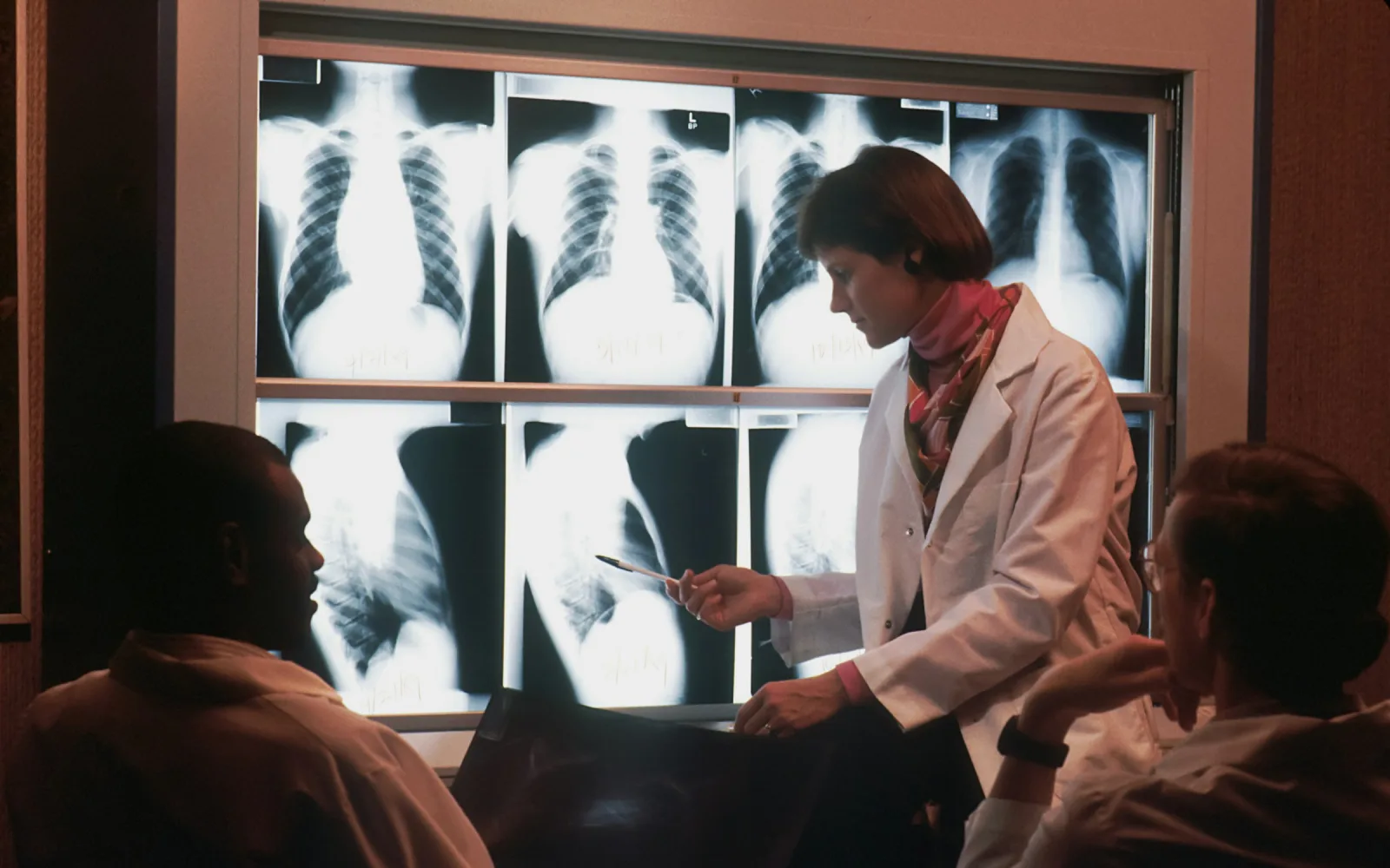
Digital X-Ray
Often the first image taken for bone pain.
Learn more
Image-Guided Pain Injection
A different procedure for pain from other causes.
Learn more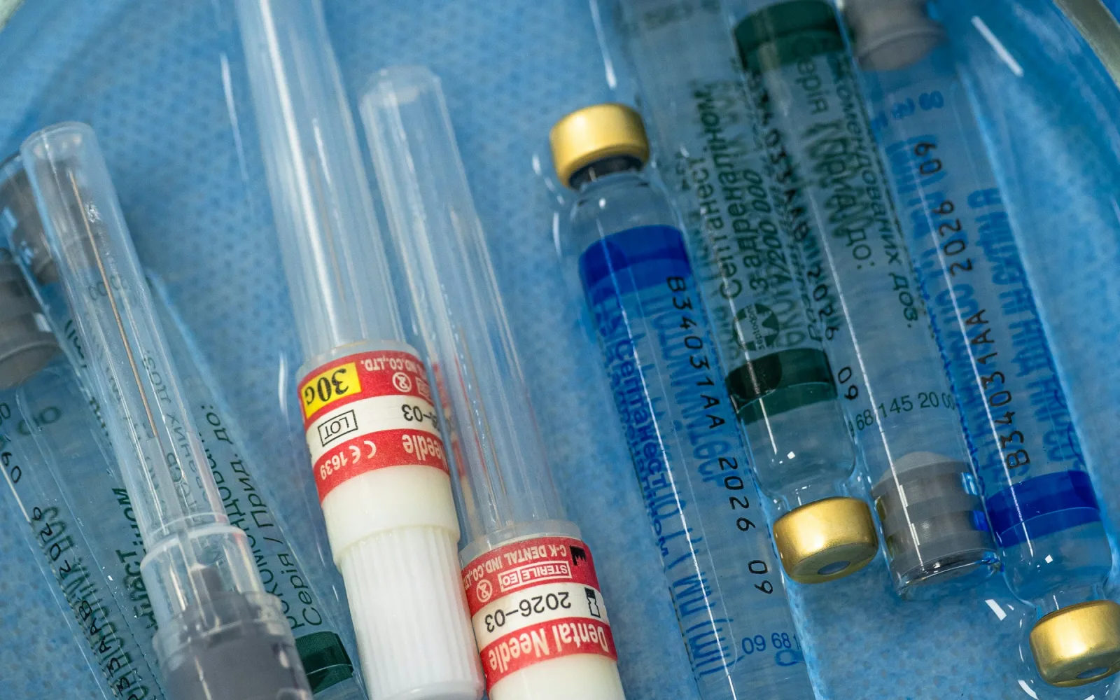
Image-Guided Biopsy
A tissue sample when the picture is not typical.
Learn more
Thyroid Nodule Ablation
Another ablation procedure, for the thyroid.
Learn moreAppointments
Ask about Osteoid Osteoma Ablation in Gomti Nagar, Lucknow
Send your prescription and any earlier reports on WhatsApp or call the centre. The team reviews them and tells you the next step.
4/12, Vijayant Khand, Gomti Nagar · 0522-3180995