Dr. Pranjali Joshi
Breast Radiologist
- MBBS (KGMU)
- MD Radiodiagnosis (Dr. RMLIMS)
- PDCC Breast Imaging (AIIMS Rishikesh)
The centre's breast radiologist is Dr. Pranjali Joshi. Ask the team when you book who will see your pictures.
Read her profile4/12, Vijayant Khand, Gomti Nagar, Lucknow Mon–Sat 9 AM–8 PM · Sun 10 AM–2 PM
Advance Breast Care • Mammography
A mammography test is a low-dose X-ray of the breast. We do screening mammograms for women with no symptoms and diagnostic mammograms for a lump or other change, at our centre in Vijayant Khand.
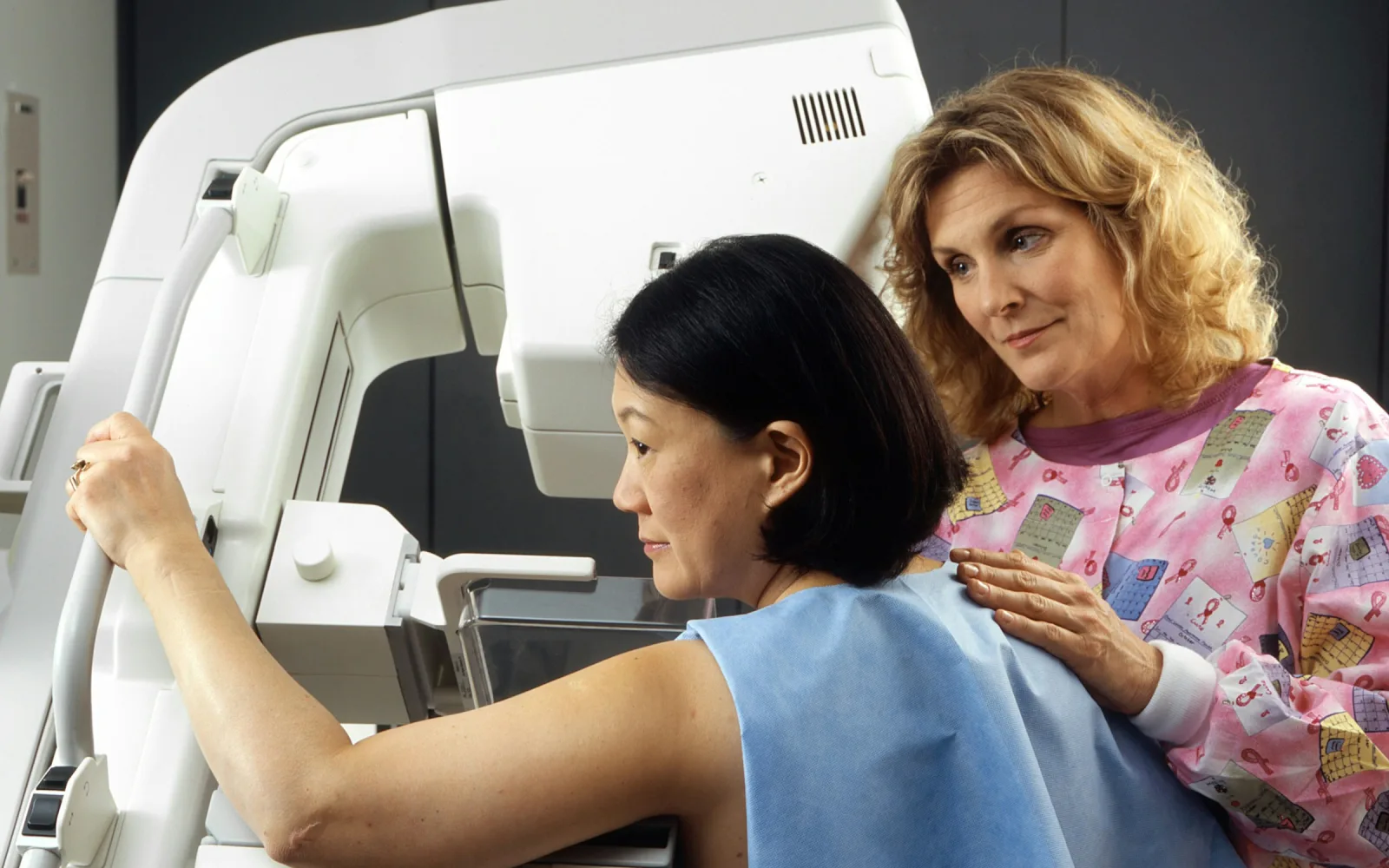
A screening mammogram is for women with no breast symptoms, and a diagnostic mammogram is for a symptom or a finding that needs looking into. The test itself is the same. The question, and the number of pictures, are different.
Tell the team which one you need when you book. If you notice a lump or other change at any point, say so at the desk, and the radiologist decides whether to add pictures to your visit.
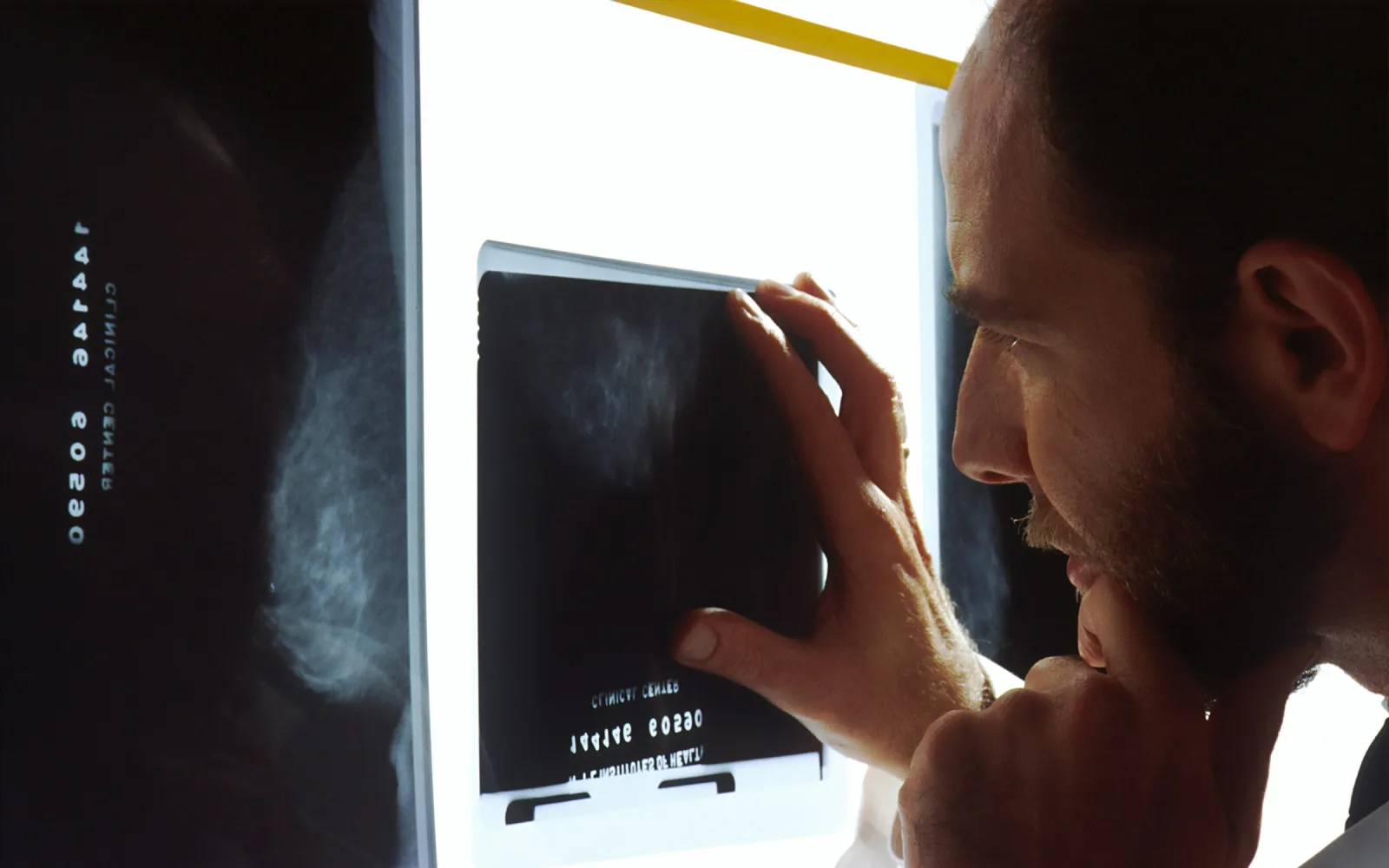
A mammogram shows the inner pattern of the breast, including small lumps, areas of distorted tissue and tiny calcium specks called calcifications. Some of these are too small to be felt, which is why mammograms are used for screening.
It has limits. Dense breast tissue can hide small changes, and a mammogram cannot tell a fluid-filled cyst from a solid lump. A lump you can feel still needs an examination and often a breast ultrasound, even when the mammogram is normal.
Many of the changes picked up on a mammogram turn out to be harmless. The radiologist's task is to separate those from the few that need more tests.
A standard mammogram takes flat 2D pictures. 3D mammography takes the pictures in thin layers, which can help in dense breasts or when a closer look is needed. Both involve the same compression and the same preparation.
Your doctor decides which suits you. If your prescription only says "mammography", the team will confirm which one is meant when you book.
Each breast is pressed between two plates for a few seconds. The pressure is uncomfortable for many women and painful for some, so say so if it is too much. Flattening the tissue gives a clearer picture from a lower dose. The radiation dose is low, and the doctor who advises the test weighs it against the benefit.
Being called back after a screening mammogram is common, and most women who are called back turn out not to have cancer.
The usual reasons are that a picture was not clear enough, that tissue overlapped, or that one area needs a closer look. The next step is usually extra mammogram views, a breast ultrasound of the area, or both. A needle test is advised only if those still leave a question.
Bring your earlier mammograms to every visit. Comparing with old pictures often settles whether an area is new or has been there for years.
Message or call the centre. Say whether this is screening or for a symptom, and mention implants, pregnancy and any earlier mammograms.
You are asked about symptoms, earlier breast tests or surgery, family history and, if you still have periods, where you are in your cycle.
You undress above the waist and put on a gown. Each breast is placed on the platform, gently drawn forward and pressed by a clear plate.
You hold still and hold your breath for a few seconds for each picture. A screening mammogram is usually two views of each breast. A diagnostic one may include more.
The team checks that the pictures are clear enough. If an area needs another view, you may be asked to wait while it is taken.
The radiologist compares your pictures with earlier ones and writes the report. The team will tell you when and how you will receive it.
Before your visit
Please bring
After your visit
A mammogram is not an emergency service. Do not hold off for an appointment if you have:
Dr. Pranjali Joshi
Breast Radiologist
The centre's breast radiologist is Dr. Pranjali Joshi. Ask the team when you book who will see your pictures.
Read her profileOur centre at 4/12, Vijayant Khand, near Kathauta Chauraha, does screening and diagnostic mammography, including 3D. Message the team on WhatsApp at +91 70809 82920 or call 0522-3180995, and say whether the mammogram is for screening or a symptom.
A prescription helps because it states the reason for the test. If you do not have one, message the team first. They will tell you what is possible, and a doctor may need to see you first if you have a symptom.
Many women find it uncomfortable and some find it painful, though the pressure lasts only a few seconds for each picture. Breasts that are tender before a period feel it more. Tell the team if it is too much, and they will adjust your position.
Usually yes. Tell the team when you book. Extra pictures are often taken to see all the breast tissue, and the plates press with care around the implant. Your doctor decides whether a mammogram is the right test for you.
Deodorants, antiperspirants and powders can contain particles that show up as small bright specks on the images. These can be mistaken for calcifications, which are tiny calcium deposits in the breast. Wash the area before you come, or tell the team if you forgot.
Not on its own. A mammogram can show that a lump looks harmless, uncertain or suspicious, but only a needle test on the tissue can confirm what it is. See breast lump evaluation for how a lump is assessed.
The gap depends on the reason for the test, your age and your risk, so your doctor will advise you. After a screening mammogram, ask your doctor when the next one is due and note the date.
Related services
A mammogram that shows the breast in thin layers.
Learn more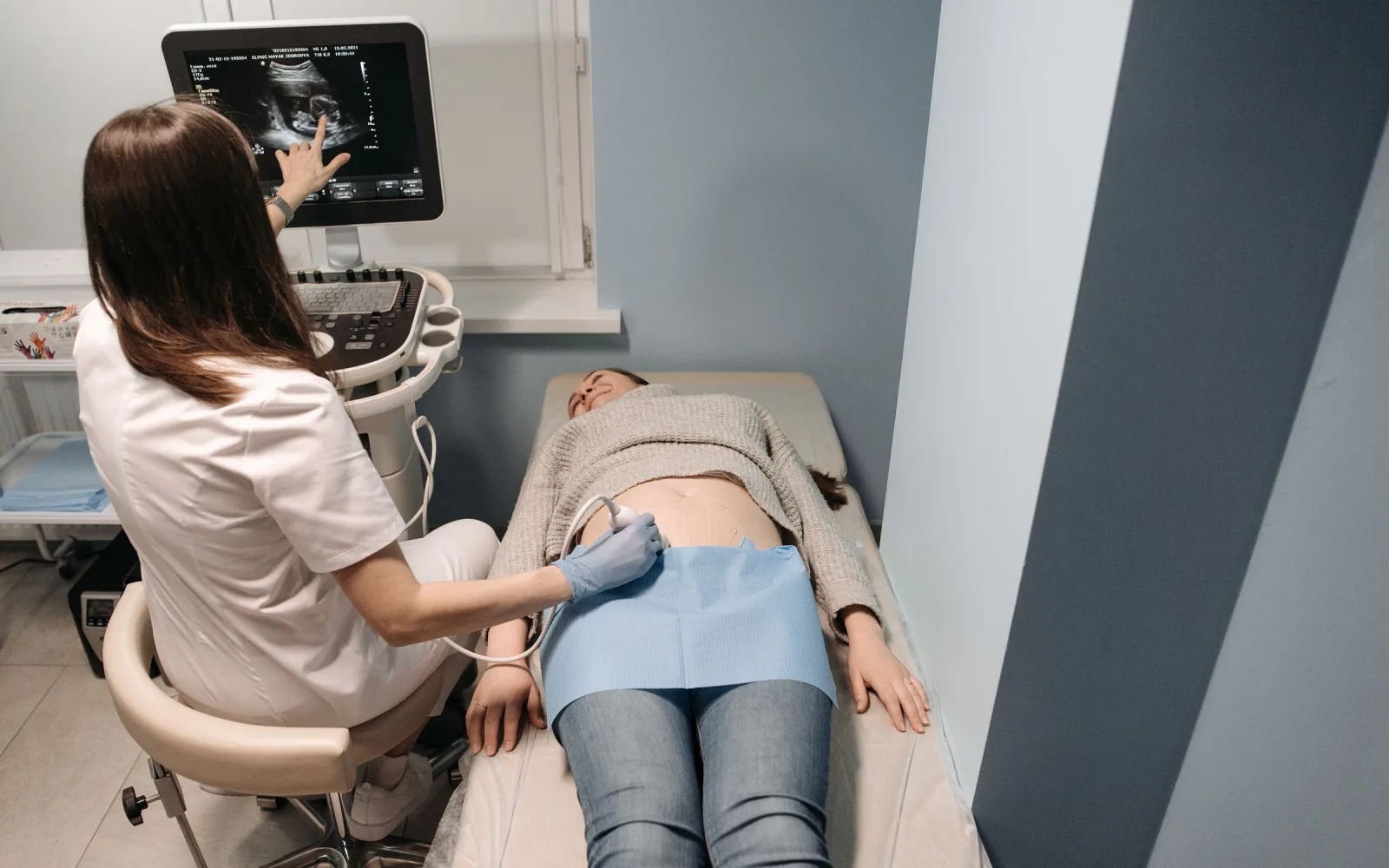
Often added to a mammogram for a lump or dense tissue.
Learn more
Who should think about a screening mammogram, and when.
Learn more
What dense breasts mean, and the extra imaging that may be advised.
Learn moreAppointments
Message us on WhatsApp or call the centre. Tell us the test you need and your preferred day, and the team will confirm the details.
4/12, Vijayant Khand, Gomti Nagar · 0522-3180995