Dr. Pranjali Joshi
Breast Radiologist
- MBBS (KGMU)
- MD Radiodiagnosis (Dr. RMLIMS)
- PDCC Breast Imaging (AIIMS Rishikesh)
Dr. Pranjali Joshi, the centre's breast radiologist, holds a PDCC in Breast Imaging from AIIMS Rishikesh.
Read her profile4/12, Vijayant Khand, Gomti Nagar, Lucknow Mon–Sat 9 AM–8 PM · Sun 10 AM–2 PM
Advance Breast Care • Breast Ultrasound
A breast ultrasound uses sound waves, not radiation, to look at a lump, dense breast tissue or a change seen on a mammogram. We do it at our centre in Vijayant Khand, Gomti Nagar.
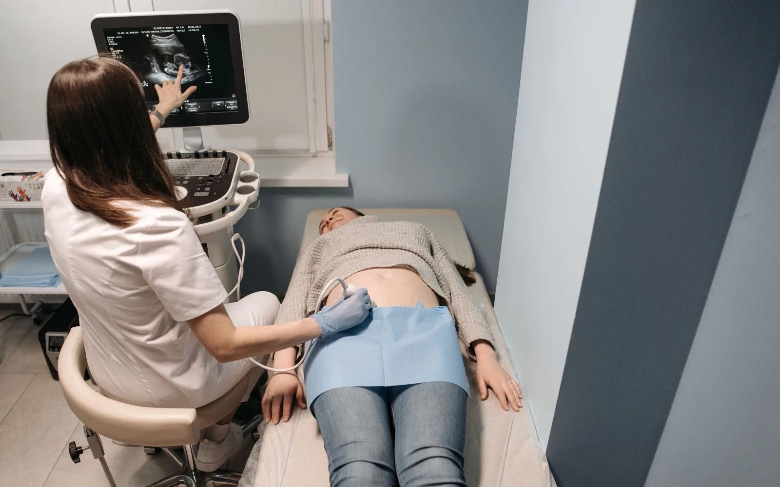
A breast ultrasound adds something a mammogram cannot give: whether a lump is solid or filled with fluid. A small probe sends sound waves into the breast and the echoes build a live picture, as in any ultrasound scan.
Cysts, which are fluid-filled sacs, look different from solid lumps such as a fibroadenoma, a common harmless lump in younger women. The radiologist can also measure a lump, describe its shape and edges, and check the lymph nodes under the arm, which are small glands that filter fluid. Underarm swelling has its own page.
When a needle test is advised, ultrasound is also the usual way to guide the needle to the right spot. Breast FNAC and breast biopsy are explained on their own pages.
Doctors advise a breast ultrasound for a lump or a spot of pain, for dense breasts, for younger women, and to look at something seen on a mammogram.
Each test sees what the other can miss, so doctors often use both. A mammogram is better at tiny calcium specks and at the overall pattern of the breast. An ultrasound is better at separating a cyst from a solid lump and at showing lumps within dense tissue.
For routine screening of women with no symptoms, doctors usually rely on mammography and add ultrasound when tissue is dense or an area needs a closer look.
A breast ultrasound cannot give a final diagnosis on its own. It can show that a lump looks harmless, uncertain or suspicious, but only a sample of the tissue can confirm what it is. It is also poor at showing the tiny calcium specks that a mammogram picks up.
If the scan looks clear but you can still feel a lump, tell your doctor. What you feel and what a doctor finds on examination matter as much as the picture.
Message or call the centre with your prescription. Tell the team where the lump or symptom is, so the right area is scanned.
You change into a gown with the chest uncovered. Hand over earlier mammograms and reports for the radiologist to compare.
You lie on your back, sometimes turned slightly to one side, with the arm on that side raised above your head. Gel is spread on the skin and the probe is moved over the breast and under the arm. You may feel light pressure.
If you have a lump, you will be asked to point to it. The radiologist scans that area with care and may measure it or take extra pictures.
The radiologist prepares the report. The team tells you when and how it reaches you, and you take it to the doctor who sent you.
Before your visit
Please bring
After your visit
A scan booking is not an emergency service. See a doctor promptly if you notice:
Dr. Pranjali Joshi
Breast Radiologist
Dr. Pranjali Joshi, the centre's breast radiologist, holds a PDCC in Breast Imaging from AIIMS Rishikesh.
Read her profileIt depends on your age, your symptoms and what your doctor is looking for. For a lump in a younger woman, ultrasound may be enough to begin with. For screening, or in older women, a mammogram is usually part of the plan. Your doctor will say which, and sometimes both are done.
Usually not. The probe presses lightly on the skin through a layer of gel. A lump or area that is already tender may feel sore when pressed. Tell the person scanning you if anything hurts.
Yes. It uses sound waves, so there is no radiation. It is often the first test when a lump is found during pregnancy or while breastfeeding. Tell the team, so the scan is planned with that in mind.
Not by itself. It shows whether a lump is a cyst or solid, and whether its shape and edges look harmless or need a closer look. A final answer comes from a needle test on the tissue, when one is needed.
It is another name for breast ultrasound, and the one you are likely to hear in India. The test is the same: sound waves and a handheld probe, with no radiation.
Yes. The radiologist can scan the lymph nodes under the arm during a breast ultrasound. A node that looks unusual may need a closer look, which depends on the whole picture.
Yes, it is safe at any point in the cycle. Breasts can be tender or lumpy just before and during a period, so the scan may be more comfortable in the week after. A new lump should not wait for the right week.
Come to 4/12, Vijayant Khand, near Kathauta Chauraha. Message the team on WhatsApp at +91 70809 82920 with your prescription, or call 0522-3180995. You can reach us from Indira Nagar, Gomti Nagar Extension and Faizabad Road.
Related services
How a lump is assessed, step by step.
Learn more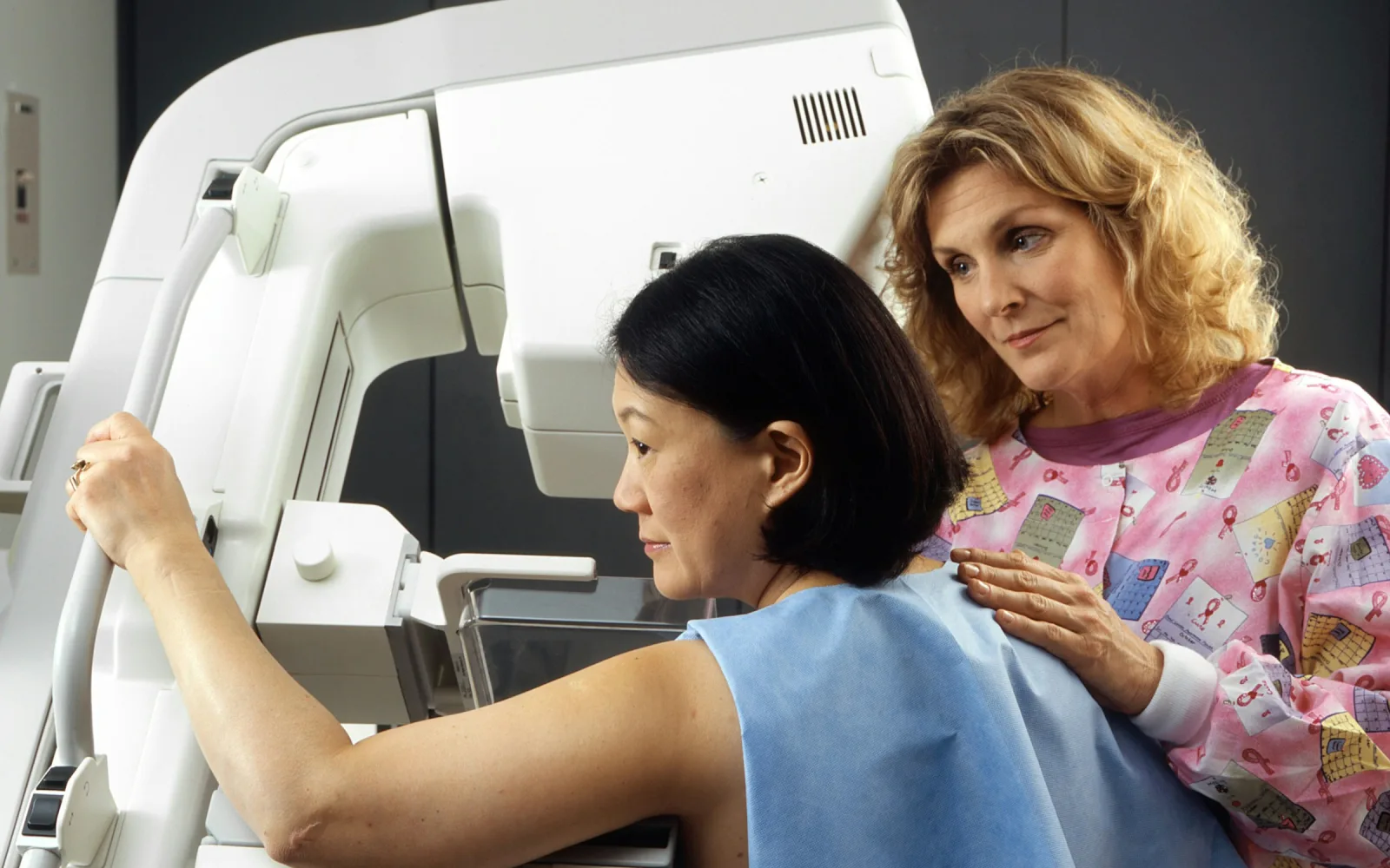
The X-ray test that ultrasound often accompanies.
Learn more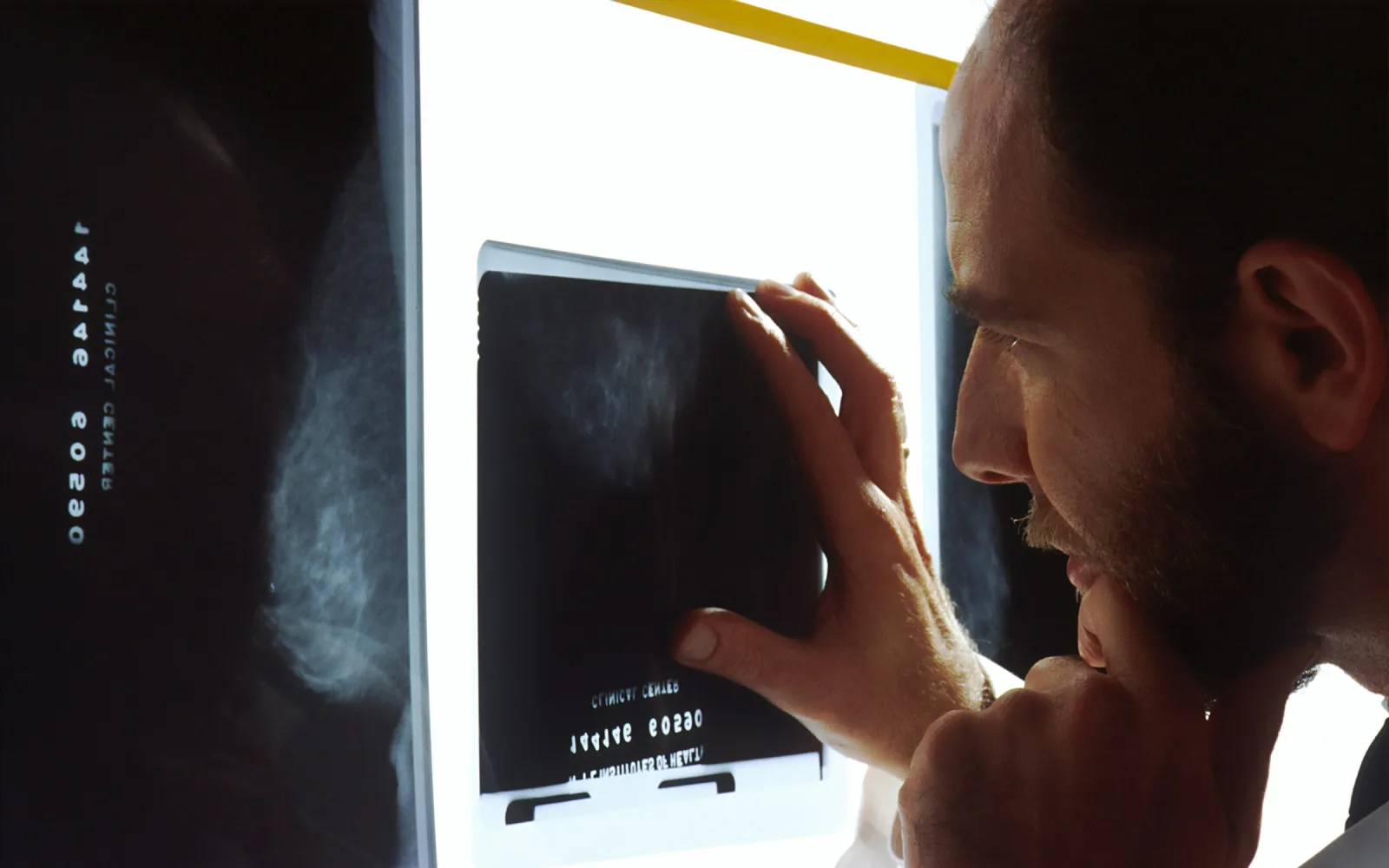
Why ultrasound may be added for dense tissue.
Learn more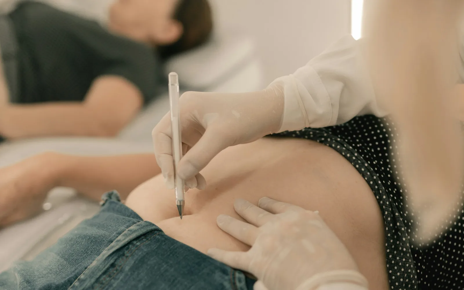
Thin-needle cell sampling from a breast lump, and when a biopsy is needed instead.
Learn moreAppointments
Message us on WhatsApp or call the centre. Tell us the test you need and your preferred day, and the team will confirm the details.
4/12, Vijayant Khand, Gomti Nagar · 0522-3180995