
Advance Breast Care • 3D Imaging

3D Mammography in Gomti Nagar, Lucknow

A 3D mammogram, also called breast tomosynthesis, is a low-dose breast X-ray that shows the breast in thin layers. It is done at our centre in Vijayant Khand, Gomti Nagar.

Call
  • Vijayant Khand, Gomti Nagar, Lucknow
  • Mon to Sat 9 AM to 8 PM, Sun 10 AM to 2 PM
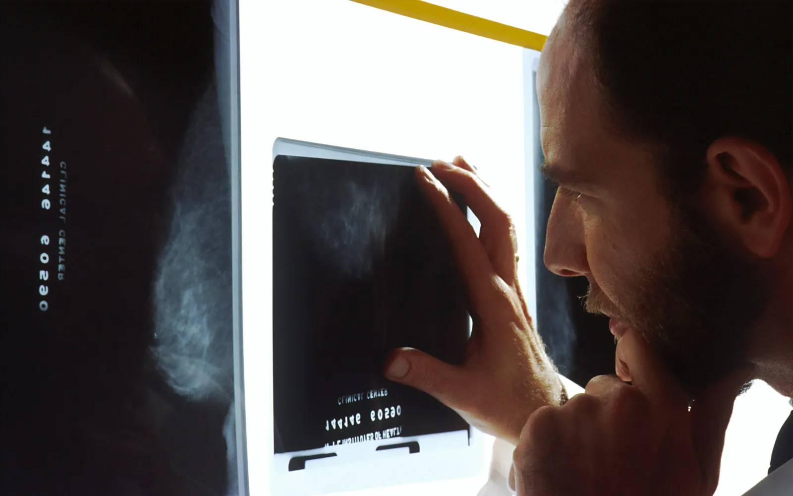
Radiologist examining mammogram films on a light box
Test
Low-dose X-ray, read in layers
Compression
A few seconds per picture
On the day
No deodorant or talc
Bring
Earlier mammograms and reports

What a 3D mammogram is

A 3D mammogram, or digital breast tomosynthesis, is a mammogram taken in a short sweep. The machine moves in an arc over the pressed breast and takes many low-dose images. A computer then rebuilds them into very thin slices.

The radiologist scrolls through the slices, much as you would turn the pages of a book one at a time. Tissue that lies on top of other tissue can be looked at layer by layer, instead of as one stacked picture.

It is still a mammogram. It uses a low dose of radiation, needs the same compression and the same preparation, and is read beside your earlier mammograms. We do 3D mammography in Lucknow for screening and for a closer look at a finding, whenever your doctor advises it.

How 3D differs from a 2D mammogram

A 2D mammogram gives one flat picture for each view of the breast, so every layer of tissue is stacked on the others. Think of looking down at a closed book: you see the cover, not the pages.

That stacking is the weakness. Normal tissue can overlap and look like a lump, or a small change can sit hidden behind other tissue. Both can lead to a call-back for more pictures.

A 3D mammogram opens the book. Because each layer can be seen on its own, overlap matters less. It does not find every change, and it does not replace an ultrasound for a lump. No breast test is perfect, which is why a symptom is always checked, whatever the scan shows.

Who may benefit from 3D mammography

Dense breasts are one of the main reasons doctors consider 3D. Dense tissue looks white on a mammogram, and so do many changes, so separating the layers can help. Dense breast imaging explains what density means and what else may be added.

  • Dense breasts: the radiologist may prefer layers to a single stacked picture.
  • A call-back after a 2D mammogram: a closer look at one area, to see whether it is a real change or overlapping tissue.
  • Follow-up of an earlier finding: the same area looked at again, in layers.
  • Screening, when your doctor advises it: breast cancer screening can use either 2D or 3D.

Many women do not need 3D, and a 2D mammogram remains a sound test. 3D is not a reason to delay a mammogram, and it never takes the place of examining a lump. For a lump, a breast ultrasound is usually part of the plan.

Radiation and compression: what to expect

The radiation dose is low. A 3D mammogram uses radiation, as a 2D one does, and the exact dose depends on how the pictures are taken. The doctor advising the test weighs that small dose against what the test can show. Tell the team if you are, or might be, pregnant.

Compression is what most women worry about. The breast is pressed between two plates for a few seconds while each picture is taken. This spreads the tissue so it can be seen, holds the breast still and allows a lower dose.

It can be uncomfortable, and for some women it hurts. Tell the team if it is too much and they will adjust. Breasts are often more tender in the days before a period, so many women prefer to book for the week after.

2D and 3D mammography at a glance

Both are mammograms read by a radiologist. This table shows where they differ.

Pictures

2D mammogram
One flat image for each view of the breast.
3D mammogram
Many low-dose images from different angles, rebuilt into thin slices.

Reading the images

2D mammogram
The radiologist studies the flat views and compares both breasts.
3D mammogram
The radiologist also scrolls through the slices, layer by layer.

Compression

2D mammogram
Yes, for a few seconds for each picture.
3D mammogram
Yes, in the same way.

Radiation

2D mammogram
A low dose.
3D mammogram
A low dose. It can be a little higher than 2D alone, depending on how the pictures are taken.

When it may be chosen

2D mammogram
Routine screening, and follow-up where 2D answers the question.
3D mammogram
Dense breasts, a closer look at a finding, or when your doctor asks for it.

Your doctor will advise which one you need. If you are unsure, send us the prescription and the team will confirm what has been asked for.

What happens during a 3D mammogram

  1. 1

    Booking

    Message or call the centre. Say if you have a lump or other symptom, breast implants or earlier mammograms, so the team can plan the right appointment.

  2. 2

    Changing

    You take off your top and any necklace, and put on a gown. Your skirt or trousers can stay on.

  3. 3

    Positioning

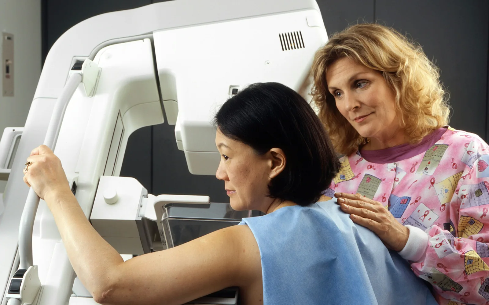
    Each breast is placed on a flat platform and a clear plate lowers to press it. The person taking the pictures adjusts your position and may ask you to lean or turn a little.

  4. 4

    The sweep

    You hold still and hold your breath for a few seconds while the machine moves over the breast. There are usually two views of each breast, one from above and one at an angle from the side.

  5. 5

    Review

    The radiologist looks at the slices beside any earlier mammograms. Sometimes an extra picture is asked for before you leave. The team will tell you when and how the report reaches you.

Preparation and aftercare

Before your visit

  • Prepare as for any mammogram: no deodorant, talc or cream on the breasts and underarms that day. The mammography page explains why.
  • Show your prescription when you book, or say you would like 3D, so the team can confirm the right appointment.
  • Wear a top you can take off easily, with a skirt or trousers rather than a dress.
  • Tell the team if you are pregnant or think you might be, or if you have breast implants.

Please bring

  • Your doctor's prescription or referral
  • Earlier mammograms (films, a disc or files) and their reports
  • Any breast ultrasound or biopsy reports

After your visit

  • Carry on with your day. You need no recovery time.
  • Your breasts may feel tender for a short while after the compression. This usually settles by itself.
  • Keep the report and the images. They are what the next radiologist compares against.
  • Take the report to the doctor who sent you. A normal report does not replace examining a new lump.

A new change should not wait for a scan date

See a doctor soon, without waiting for a mammogram appointment, if you notice:

  • A new lump or thickened area in the breast or under the arm.
  • Blood-stained fluid from the nipple, or fluid from one nipple without squeezing.
  • Dimpling of the skin, or a nipple that has recently turned inwards.
  • A red, hot, swollen and painful breast, especially with a fever.

Meet our radiologist

Dr. Pranjali Joshi, Breast Radiologist at Lucknow Imaging Centre

Dr. Pranjali Joshi

Breast Radiologist

  • MBBS (KGMU)
  • MD Radiodiagnosis (Dr. RMLIMS)
  • PDCC Breast Imaging (AIIMS Rishikesh)

The centre's breast radiologist, Dr. Pranjali Joshi, has postdoctoral training in breast imaging from AIIMS Rishikesh.

Read her profile
3D Mammography: common questions

Is 3D mammography better than a 2D mammogram?

It is different rather than simply better. 3D separates overlapping tissue, which helps some women, especially those with dense breasts. A 2D mammogram is still a sound test, and many women need nothing more. Your doctor chooses using your age, breast density, symptoms and earlier results.

Does a 3D mammogram hurt more than a normal one?

Not usually. Compression works in the same way as for a 2D mammogram and lasts a few seconds for each picture. If it becomes too much, say so and the team will adjust your position.

Is the radiation from 3D mammography safe?

The dose is low, though it can be a little higher than for a 2D mammogram alone. Your doctor weighs that small dose against what the test can show. If you are pregnant or think you might be, tell the team before any pictures are taken. Mammograms are usually avoided in pregnancy unless the benefit is clear.

Do I need a 3D mammogram if I have dense breasts?

Not always. 3D can help the radiologist separate overlapping dense tissue, but some people with dense breasts need only a 2D mammogram, and others have a breast ultrasound added. Your doctor will advise on the combination that suits your age, risk and symptoms.

Can 3D mammography replace a breast ultrasound?

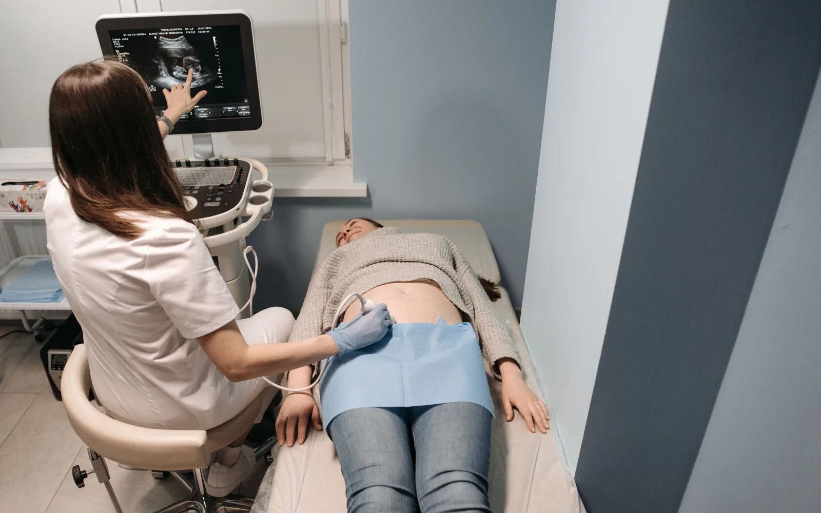
No, because they answer different questions. A mammogram, 2D or 3D, shows the pattern of the whole breast and tiny calcium specks. An ultrasound shows whether a lump is a fluid-filled cyst or a solid lump. For a lump, doctors often want both.

Where can I get a 3D mammogram near Kathauta Chauraha?

We do 3D mammography at 4/12, Vijayant Khand, near Kathauta Chauraha, Gomti Nagar, Lucknow. Message the team on WhatsApp at +91 70809 82920 or call 0522-3180995, and they will confirm that 3D is what your doctor has asked for.

How long will I wait for the report?

The team will tell you when you book or on the day, so you can plan your follow-up with your doctor. The radiologist prepares the report and, if you bring earlier mammograms, compares your new pictures with them first.

Last updated 12 October 2026.

This page gives general information to help you prepare. It does not replace advice from your own doctor, who knows your history and examination findings.

Appointments

Book 3D Mammography in Gomti Nagar, Lucknow

Message us on WhatsApp or call the centre. Tell us the test you need and your preferred day, and the team will confirm the details.

4/12, Vijayant Khand, Gomti Nagar · 0522-3180995

Reception at Lucknow Imaging Centre, Gomti Nagar