Breast Procedure • Gomti Nagar

Breast Biopsy in Gomti Nagar, Lucknow

A breast biopsy in Lucknow takes a few small samples of tissue with a needle, guided by imaging, so a pathologist can say what a lump or abnormal area is. Here is how it works and what the report means.

Call
  • Vijayant Khand, Gomti Nagar, Lucknow
  • Mon to Sat 9 AM to 8 PM, Sun 10 AM to 2 PM
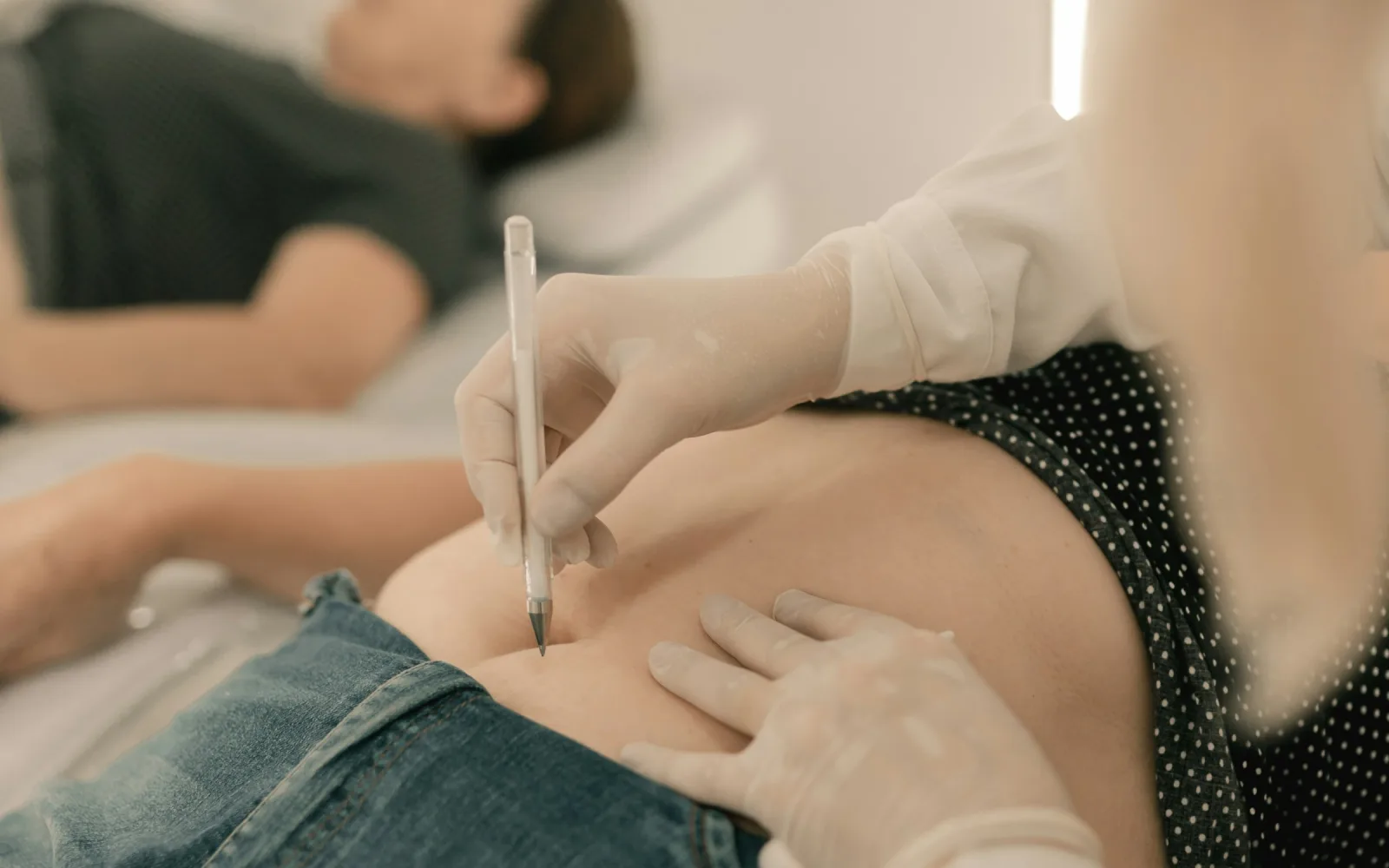
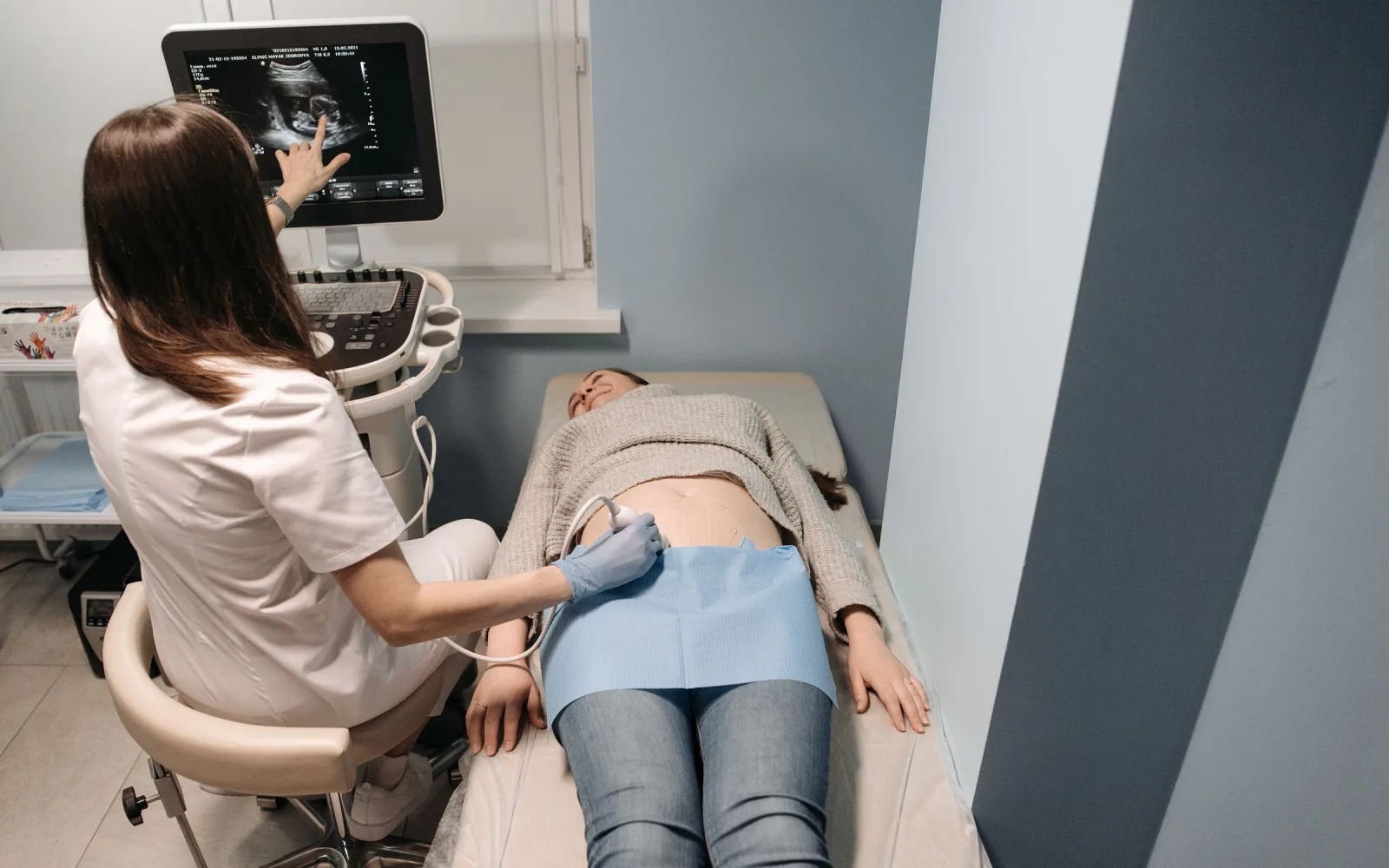
Gloved clinician marking the skin before an image-guided needle procedure
Sample
A few thin tissue cores
Numbing
Local anaesthetic first
Guidance
Imaging, usually ultrasound
Result
Pathology report, then review

What is a breast biopsy?

A breast biopsy removes a small amount of tissue so that it can be examined under a microscope. The usual method is a core needle biopsy. A hollow needle takes several thin cores of tissue, and a pathologist studies them to say what the lump or area is made of. Biopsies of other organs are covered on the general biopsy page. This page is about the breast.

Doctors advise it when a scan or an examination shows something that needs a firm answer. Examples are a lump that looks suspicious, a cluster of tiny calcium specks on a mammogram, or a lump where a breast FNAC result was unclear. A biopsy is advised to find out what an area is, and the answer can be harmless or not. Most breast changes that lead to a biopsy turn out not to be cancer.

How does imaging guide the needle?

Imaging shows the doctor where the needle is, so it reaches the exact spot. Most lumps that show on a breast ultrasound are sampled with the needle visible on the screen as it goes in. The same idea is used in other organs, as described on the USG-guided biopsy page.

Some findings, such as tiny calcium deposits, show only on a mammogram. They may need mammogram-based guidance, called stereotactic biopsy, instead. The team reviews your scans and confirms which method suits your finding, and whether it can be arranged at the centre or needs another facility.

If the spot may need to be found again, for example before chemotherapy or surgery, the doctor can leave a tiny clip marker at the biopsy site. It is not needed after every biopsy. When surgery is planned for something that cannot be felt, wire localisation helps the surgeon find it.

What the biopsy report means in plain words

The tissue is processed in a laboratory, cut into very thin slices and studied by a pathologist. The report describes what the cells and the tissue pattern look like. The radiologist and your treating doctor then compare the report with your scans to check that the two fit together. If they do not, a repeat biopsy or another test may be advised.

A pathology result is meant to be discussed with a doctor, not read alone as a line on paper. The team will tell you when the report is expected and who goes through it with you. Plan a visit with your treating doctor to discuss it, and bring someone with you if that helps.

Benefits, limits and risks

A biopsy gives a firm answer about what a lump or area is. When the finding is benign, it can spare you an operation. It is not perfect, though. The needle can miss part of a lesion, and a small sample may not show the whole picture. That is why the result is read with the scan, and why a repeat sample or follow-up imaging is sometimes advised.

Common effects are bruising, soreness and a little bleeding at the site. The bruise can look large and fade over a week or two without being a problem. Less often, a collection of blood forms in the breast, or the site becomes infected. Serious complications are rare.

Tell the team about blood thinners, aspirin, bleeding problems, allergies and any chance of pregnancy. Do not stop prescribed medicines unless your doctor tells you to. The doctor may ask for a blood count or other blood tests beforehand. The team confirms suitability and who carries out the biopsy for each case.

Common words in a biopsy report

These words appear often. The table explains them in plain language. It is not a diagnosis, and your doctor will explain what your own report means.

Benign

In plain words
No cancer cells were seen. Examples are a fibroadenoma (a common harmless lump), a cyst, or fibrocystic change (lumpy but harmless breast tissue).
What may follow
Often reassurance, or a repeat scan after a few months, as your doctor advises.

Atypical or high-risk lesion

In plain words
Some cells look unusual but are not cancer.
What may follow
More imaging or sampling, and sometimes removal of the area, to be sure nothing more is present.

In situ, for example DCIS

In plain words
Abnormal cells are present but stay inside the milk ducts. They have not grown into the surrounding breast.
What may follow
Review by a breast surgeon and the cancer team.

Invasive cancer

In plain words
Cancer cells have grown into the breast tissue. The report often adds the type, grade and hormone receptor results.
What may follow
Planning with a breast surgeon or cancer specialist, who will explain each step.

Insufficient or non-diagnostic

In plain words
Too little tissue, or tissue that does not explain the scan finding.
What may follow
The biopsy may be repeated or another test used.

What happens on the day

  1. 1

    Booking and review

    Send your doctor's note, scans and earlier reports. The team reviews them, confirms that a biopsy is appropriate and which guidance method is needed, and tells you how to prepare.

  2. 2

    Check and consent

    The doctor checks your history, medicines and allergies, shows you the finding on the scan and explains the procedure. You are asked to sign a consent form. Ask any question before you do.

  3. 3

    Numbing

    The skin is cleaned and local anaesthetic is injected. It stings for a few seconds, and then the area goes numb.

  4. 4

    Taking the samples

    The needle is placed under imaging and a few cores are taken. You may hear a clicking sound and feel pressure. Most people do not feel sharp pain, but tell the doctor if you do. A marker is left at this stage only if it was planned.

  5. 5

    Pressure and dressing

    Firm pressure is held on the site for several minutes to limit bruising. A dressing covers the spot, and the team explains aftercare.

  6. 6

    Results

    The tissue goes to the laboratory. The team tells you when to expect the report, and you arrange a visit with your doctor to go through it.

Preparation and aftercare

Before your visit

  • Most people can eat and drink normally beforehand. If the team asks you to fast, follow their advice.
  • Tell the team about blood thinners, aspirin, herbal supplements and any bleeding problem. Do not stop any of them unless your doctor tells you to.
  • Mention allergies, especially to local anaesthetic, plasters or dressings.
  • Choose a loose top that opens at the front.
  • Arrange for someone to come with you, and plan a quiet rest of the day.

Please bring

  • Your doctor's note or referral
  • All earlier breast scans, with the images as well as the reports
  • Any earlier FNAC or biopsy report
  • A list of your medicines

After your visit

  • Keep the dressing in place and dry for the first day, or as the team advises.
  • A firm, supportive bra may feel more comfortable for a day or two.
  • Avoid heavy lifting, vigorous exercise and swimming for a day or two, or as advised.
  • Soreness and bruising are common and settle over days. If you need a painkiller, ask the team which one suits you.
  • Book a time with your treating doctor to go through the report.

Warning signs after a biopsy

A bruise and soreness are expected. Contact the team or your doctor the same day, or go to the nearest emergency department if you cannot reach them, if you have:

  • Bleeding that comes through the dressing and keeps going after ten minutes of firm pressure.
  • A breast that quickly becomes larger, tight, hard or very painful.
  • Fever, spreading redness, warmth or pus from the site.
  • Pain that gets worse after the first day instead of easing.
  • Breathlessness, chest pain or a feeling that you may faint, which need urgent care.

Meet our radiologist

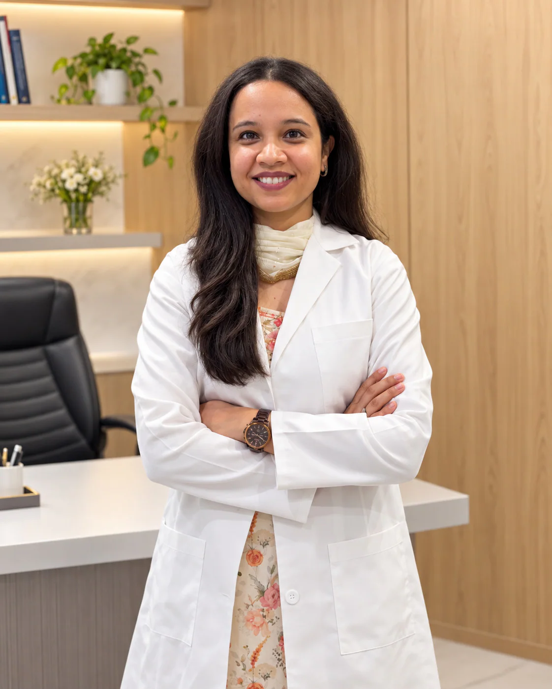
Dr. Pranjali Joshi, Breast Radiologist at Lucknow Imaging Centre

Dr. Pranjali Joshi

Breast Radiologist

  • MBBS (KGMU)
  • MD Radiodiagnosis (Dr. RMLIMS)
  • PDCC Breast Imaging (AIIMS Rishikesh)

Dr. Pranjali Joshi is the centre's breast radiologist. The team reviews your scans first, then confirms for your case whether a biopsy is suitable and who will carry it out.

Read her profile
Breast Biopsy: common questions

Is a breast biopsy painful?

The numbing injection stings briefly. After that most people feel pressure and hear a click rather than feel sharp pain. Soreness afterwards is common and eases over a few days. If you feel pain during the procedure, say so, and the doctor can add more anaesthetic.

Does a breast biopsy mean I have cancer?

Not by itself. A biopsy is a test to find out what a lump or area is, and the answer can be harmless or not. Most breast changes that lead to a biopsy turn out not to be cancer. The test is advised so that a clear answer replaces uncertainty.

Can a needle biopsy make breast cancer spread?

There is no good evidence that a needle biopsy makes breast cancer spread. Biopsy is the standard way to get a diagnosis before treatment is planned. Ask your doctor if this worry stays with you.

How long does a breast biopsy take?

The needle part is usually short, but the visit is longer because of checks, numbing, pressure afterwards and a short rest. The team will tell you how long to plan for when you book.

How and when are the breast biopsy results explained?

The laboratory needs time to process the tissue, and the timing depends on the tests ordered. The team will tell you when the report is expected. A result is best discussed in person with your doctor, who can explain the words and the next step.

Can I go to work after a breast biopsy?

Many people can return to desk work the next day. Avoid heavy lifting and vigorous exercise for a day or two. The team will advise you for your own case, so ask before you leave.

Do I need a clip or marker after a breast biopsy?

Not always. Most biopsies end without one. A marker is added only when the doctor expects to need the exact spot again later, and the page on clip marker placement explains when that applies. The doctor will tell you beforehand if one is planned.

Where can I get a breast biopsy near Kathauta Chauraha, Gomti Nagar?

The centre is at 4/12 Vijayant Khand, near Kathauta Chauraha, Gomti Nagar. Message us on WhatsApp at +91 70809 82920 or call 0522-3180995 with your scans. The team confirms whether a biopsy suits your finding and who will carry it out.

Last updated 12 October 2026.

This page gives general information to help you prepare. It does not replace advice from your own doctor, who knows your history and examination findings.

Appointments

Ask about Breast Biopsy in Gomti Nagar, Lucknow

Send your prescription and any earlier reports on WhatsApp or call the centre. The team reviews them and tells you the next step.

4/12, Vijayant Khand, Gomti Nagar · 0522-3180995

Reception at Lucknow Imaging Centre, Gomti Nagar