Interventional Radiology • Gomti Nagar
Renal Artery Embolization for Haematuria in Lucknow
Blood in the urine always needs a doctor to look for the cause. Renal artery embolization can stop bleeding from a kidney artery in selected cases, once that cause has been looked for.
- Vijayant Khand, Gomti Nagar, Lucknow
- Mon to Sat 9 AM to 8 PM, Sun 10 AM to 2 PM
- Blood in urine
- Needs urgent assessment
- What it does
- Blocks the bleeding kidney artery
- First scan
- Ultrasound, Doppler if needed
- Where it is done
- Confirmed after case review
Blood in the urine: why it needs urgent assessment
Blood in the urine, called haematuria, always needs a doctor to find the cause. It may show as red, pink or tea-coloured urine, or be found only on a urine test. Bleeding that causes no pain is not a reason to wait, because some serious causes are silent.
See a doctor the same day if the bleeding is heavy, if you pass clots, if you cannot pass urine, or if you have fever or severe pain in the side. Go to a hospital emergency department if you feel faint or dizzy.
Common causes are urine infection, kidney or bladder stones, an enlarged prostate, and tumours of the kidney, bladder or prostate. Less often the cause is an injury, a blood vessel problem in the kidney, or bleeding after a kidney biopsy or surgery. Blood thinners and bleeding disorders can make bleeding worse or bring it out.
What is renal artery embolization?
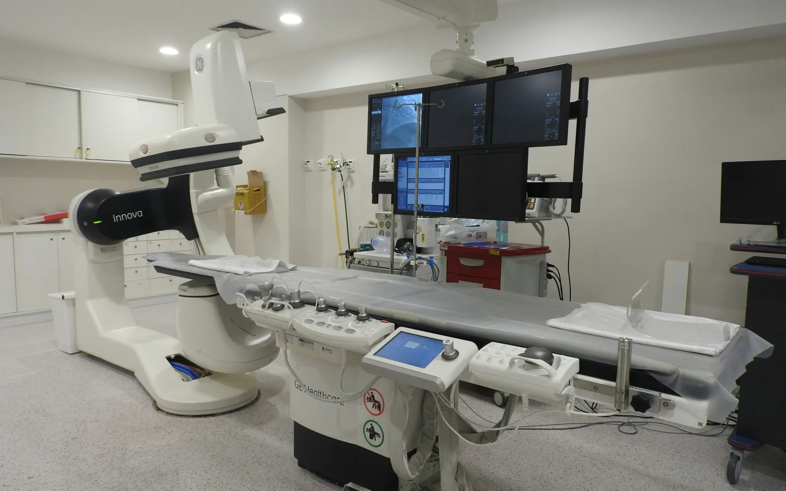
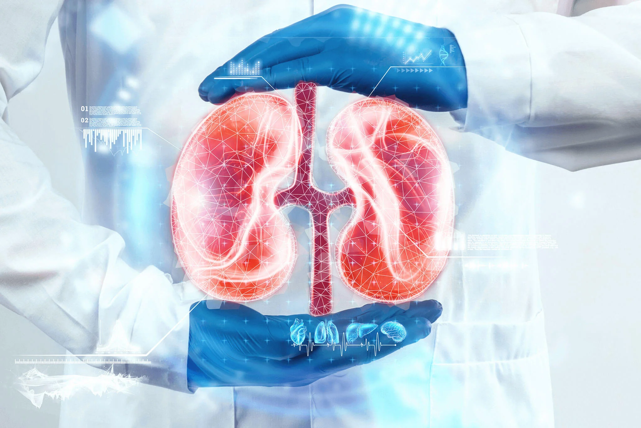
Renal artery embolization blocks a small artery inside the kidney that is bleeding. A thin catheter is passed into an artery, usually at the groin, and guided to the renal artery under live imaging. Dye shows the branch that is bleeding or abnormal.
Tiny particles, coils or other material are then released into that branch to close it. The aim is to treat only the bleeding branch and leave as much of the kidney working as possible.
It is considered for selected arterial causes. These include an injury to the kidney, bleeding after a kidney biopsy or surgery, an abnormal tangle of vessels, a small bulge in an artery wall called a pseudoaneurysm, and a bleeding kidney growth. It is not used for the commonest causes of blood in the urine, such as infection or stones, which are treated differently.
How ultrasound and Colour Doppler help
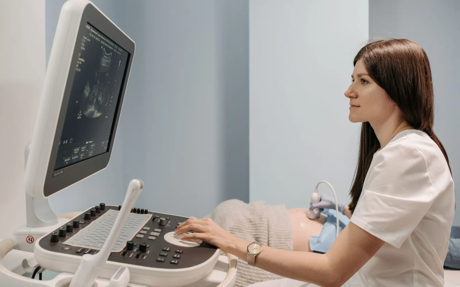
An ultrasound of the kidneys, ureters and bladder is usually the first scan. It can show stones, a blocked kidney, a growth, and clots in the bladder, without radiation or dye. That is useful when the kidneys need protecting from dye.
Colour Doppler adds blood-flow information. It can show an abnormal connection between an artery and a vein, or a small bulge in an artery, which can follow a kidney biopsy or injury. It also shows how well blood flows into the kidney.
These scans cannot always find the exact bleeding point. If bleeding continues or an arterial cause is suspected, a CT scan with dye is usually the next step, because it can show active bleeding and the artery behind it. CT and MRI are not available at the centre. Your doctor advises where to get one, and we can review the images and reports.
Other options, benefits and risks
Treatment depends on the cause. A urine infection is treated with antibiotics, stones are dealt with by a urologist, and a doctor may change medicines that add to bleeding. Urologists can also look inside the bladder with a camera, called cystoscopy, and treat prostate bleeding. Surgery to remove part or all of the kidney is another option, usually kept for bleeding that cannot otherwise be controlled.
Embolization is useful when the source is an artery and the aim is to keep the kidney, but it carries risks:
- Loss of kidney tissue: the blocked area loses its blood supply, so the kidney may work slightly less. This matters more if kidney function is already low.
- Post-embolization symptoms: pain in the side, fever, nausea and tiredness for some days.
- Dye problems: an allergic reaction, or strain on the kidneys, especially if kidney function is already poor or you are dehydrated or diabetic.
- Treatment reaching the wrong place: material can reach a branch it was not meant to block.
- Puncture site: bruising, bleeding or damage to the artery at the groin.
- Bleeding returning, or infection of the treated area.
No one can promise that the bleeding will stop for good. If it returns, the team and your urologist will talk about the next step.
How the decision is made, and where it is done
The urologist or nephrologist treating you decides together with the interventional radiologist. They look at the scans, the amount of blood lost, kidney function and clotting, and whether a safer option exists.
A kidney function panel checks creatinine, which shows how well the kidneys clear dye and waste. A urine test checks for infection. Both are usually done before any dye is used.
Our team reviews your referral, scans and reports first, then confirms whether the procedure is carried out at the centre or elsewhere, and by whom. Heavy bleeding with dizziness or clots is dealt with at a hospital emergency department, not through a booking.
From first assessment to follow-up
-
1
Urgent assessment
A doctor examines you, checks blood pressure, haemoglobin and kidney function, and tests the urine. Heavy bleeding is managed in hospital first.
-
2
Imaging
An ultrasound is often done first, with Colour Doppler added if a blood vessel cause is suspected. A CT scan with dye is the usual next step if an arterial source is suspected, and your doctor advises where to have it.
-
3
Case review
Send us the referral, scans and reports. The team reviews them and confirms where the procedure can be carried out, and by whom.
-
4
Planning and consent
Before the day, the doctor explains the approach, the other options and the risks, and asks for your consent. Blood tests and clotting are checked.
-
5
The procedure
The catheter is guided to the kidney artery, the bleeding branch is found with dye and then blocked. You are told beforehand what sedation or anaesthesia is planned.
-
6
Recovery and follow-up
You are watched for bleeding, pain and fever, and kidney tests are repeated. Your urologist continues to look for and treat the cause.
Preparation and aftercare
Before your visit
- Keep every ultrasound, Doppler and CT report together with the images, and your urine and blood test reports.
- Note when the blood first appeared, its colour, whether you pass clots, and any pain, fever or injury.
- Tell the team about allergies, kidney disease, diabetes and all your medicines, especially blood thinners and painkillers.
- Never stop a prescribed medicine on your own. Blood thinners need a decision from the treating doctor.
- The team doing the procedure tells you how long to fast.
Please bring
- Doctor's referral and discharge or clinic notes
- Ultrasound, Doppler and CT reports and images
- Urine and blood test reports
- List of current medicines
After your visit
- Drink fluids as advised so the kidneys can clear the dye, unless you were told to limit fluids.
- Take the prescribed medicines for pain and fever. Avoid other painkillers unless the team says they are safe, as some can strain the kidneys.
- Keep the puncture site clean and dry, and avoid heavy lifting for as long as the team advises.
- Repeat the kidney tests and urine checks as advised, and keep the follow-up visit with your urologist.
Blood in the urine: do not wait
Seek urgent medical care, at a hospital emergency department if needed, for blood in the urine with any of these. After embolization, do the same if you have them.
- Heavy bleeding, clots in the urine, or being unable to pass urine.
- Passing much less urine than usual.
- Fever with shivering, or severe pain in the side or abdomen.
- Dizziness, fainting, a racing heart or looking pale.
- Swelling, bleeding or a lump at the groin, or a cold, pale or numb leg.
- Chest pain or breathlessness.
Renal Artery Embolization: common questions
Is blood in the urine always serious?
Not always, but it should always be checked. A urine infection or a stone is a common cause, yet some serious conditions cause nothing but blood in the urine. The cause is looked for with urine tests and scans.
Which scan is done first for blood in the urine?
An ultrasound of the kidneys and bladder is often the first scan. It shows stones, blockage, growths and clots without radiation or dye. Colour Doppler is added when a blood vessel problem is suspected.
Can Colour Doppler find a bleeding artery?
Sometimes. It can show an abnormal artery-to-vein connection or a small bulge in an artery, especially after a biopsy. It cannot always find the exact bleeding point, and a CT scan with dye is often needed.
Does embolization damage the kidney?
It can reduce kidney tissue in the area that is blocked. The team aims to block only the bleeding branch, so that as much kidney as possible keeps working. Kidney function is checked before and after, and the risk is higher if function is already low.
Is renal artery embolization the same as angioplasty?
No. Angioplasty and stenting open a narrowed artery, for example when it limits blood flow to the kidney or raises blood pressure. Embolization does the opposite: it closes a bleeding artery.
Can blood thinners cause blood in the urine?
They can make bleeding more likely or bring out an underlying problem, but blood in the urine on a blood thinner still needs to be investigated. Do not stop the medicine yourself. Ask the doctor who prescribed it, the same day.
Do you perform renal artery embolization in Lucknow?
The team tells you after reviewing your referral, scans and reports. Whether the procedure is carried out at the centre or elsewhere, and by whom, is confirmed for each case. Heavy bleeding needs a hospital emergency department first.
Related services
Tests and care that often go together

Appointments
Ask about Renal Artery Embolization for Haematuria in Gomti Nagar, Lucknow
Send your prescription and any earlier reports on WhatsApp or call the centre. The team reviews them and tells you the next step.
4/12, Vijayant Khand, Gomti Nagar · 0522-3180995